. 2001 Jun 5;98(12):6599-604.
doi: 10.1073/pnas.121171498. Epub 2001 May 29.

Preferential interaction of the core histone tail domains with linker DNA

Affiliations

Preferential interaction of the core histone tail domains with linker DNA

D Angelov et al. Proc Natl Acad Sci U S A. .

Abstract

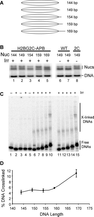
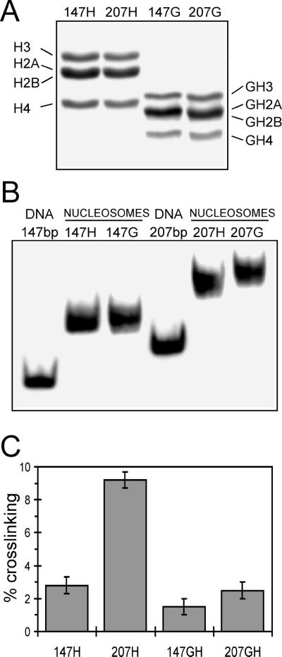
Within chromatin, the core histone tail domains play critical roles in regulating the structure and accessibility of nucleosomal DNA within the chromatin fiber. Thus, many nuclear processes are facilitated by concomitant posttranslational modification of these domains. However, elucidation of the mechanisms by which the tails mediate such processes awaits definition of tail interactions within chromatin. In this study we have investigated the primary DNA target of the majority of the tails in mononucleosomes. The results clearly show that the tails bind preferentially to "linker" DNA, outside of the DNA encompassed by the nucleosome core. These results have important implications for models of tail function within the chromatin fiber and for in vitro structural and functional studies using nucleosome core particles.


Figures

Figure 1
Figure 1
Schematic of the sample preparation and experimental protocol for studying the effect of the length of nucleosomal DNA on the crosslinking efficiency of the core histone tail domains. MNase, micrococcal nuclease; 5% Acry NP gel, native 5% polyacrylamide nucleoprotein gel.
Figure 2
Figure 2
The efficiency of histone–DNA crosslinking within nucleosomes depends on DNA length. Native H1-depleted oligosomes in buffer containing 80 mM NaCl, 0.25 mM EDTA, and 10 mM Tris⋅HCl, pH 7.5, were irradiated with a single UV laser pulse, then nucleosomes were separated on a 5% polyacrylamide gel. The nucleosome band was cut into several successive slices, the nucleoprotein complexes were electroeluted, and the percentage of crosslinked histone–DNA complexes was determined. (A) DNA length determination for the histone-DNA complexes isolated from the mononucleosome slices. The histone–DNA complexes were extensively digested with Pronase, and DNA was isolated and run on a native 5% polyacrylamide gel. Left lane 1, 1-kb DNA ladder. (B) Plot of the percent of total DNA crosslinked vs. nucleosomal DNA length. (C) As in B except that the nucleosomal DNAs were radioactively end-labeled before the irradiation step (see text). The data represent average values and standard deviations from four different experiments.
Figure 3
Figure 3
Linker-DNA length-dependent crosslinking of the core histone tails is observed in low ionic strength buffers. Nucleosomes were prepared and irradiated as described in for Fig. 2 except that the buffer contained 1 mM NaCl. (A) DNA length determination for the histone–DNA complexes isolated from the mononucleosome slices. (B) Plot of the percent of total DNA crosslinked vs. nucleosomal DNA length.
Figure 4
Figure 4
Immunoslot analysis of the reaction of antibodies to the individual core histones H2A, H2B, H3, and H4 and preimmune IgG (0) with covalent histone complexes. H1-depleted oligosomes (essentially monosomes, containing small amount of di- and trisomes, see Fig. 1) and core particles were UV laser irradiated with identical doses, and the covalent complexes were purified on CsCl gradients. A series of 5 μg and 2.5 μg of the complexes (measured as DNA) as well as of the control nonirradiated (CON) CsCl-purified samples were loaded on nitrocellulose filters, and the crosslinked histones were detected with immunopurified antibodies. The slots loaded with H1-depleted oligonucleosomes (ONS) or core particles (CPS) are indicated.
Figure 5
Figure 5
Efficiency of targeted photochemically induced crosslinking depends on nucleosomal DNA length. Nucleosomes were reconstituted with a recombinant H2B containing a single cysteine at position two in the N-terminal tail (H2B2C) and modified with APB. (A) DNA templates used for reconstitution. Nucleosomes were reconstituted with specific DNA templates ranging in length from 144 to 169 bp. (B) Nucleosomes assembled on various DNA templates. Reconstituted mixtures containing nucleosomes and unassembled (naked) DNAs were separated on a nucleoprotein gel and visualized by autoradiography of the wet gel. Lanes 1–5 contain irradiated nucleosomes reconstituted with 144-, 149-, 154-, 159-, and 169-bp templates, respectively. Lanes 6 and 7 contain nucleosomes reconstituted with completely wild-type histones that were either unirradiated or irradiated before separation on the gel, respectively. Lane 8 contains nucleosomes reconstituted with H2B2C that was not further modified with the crosslinking reagent. (C) Products of site-specific photochemical crosslinking reactions. Nucleosomes and naked DNAs were eluted from the preparative nucleoprotein gel, denatured with SDS, then subjected to SDS/PAGE to visualize protein–DNA crosslinked products. Lanes 1 and 2 contain naked 149-bp DNA either unirradiated or UV irradiated, respectively. Lanes 3 and 4 contain nucleosomes reconstituted with wild-type histones and either unirradiated or UV-irradiated, respectively. Lane 5 contains nucleosomes reconstituted with H2A2C-APB and 149-bp DNA but not irradiated. Lanes 6–10 contain UV-irradiated nucleosomes reconstituted with H2A2C-APB and the 144-, 149-, 154-, 159-, or 169-bp templates, respectively. Lanes 11–15 contain samples identical to those in lanes 6–10 but from a separate experiment. (D) Summary plot of percent DNA crosslinked vs. nucleosomal DNA length (in bp). The fraction of radiolabeled DNA fragment present in crosslinked species (see C) was determined and plotted vs. nucleosomal DNA length.
Figure 6
Figure 6
UV laser-induced crosslinking within a defined sequence nucleosome and nucleosome core particle. Either a 147- or a 207-bp 5S DNA fragment was reconstituted with either full-length or tailless recombinant core histones, then the nucleosomes were purified by sucrose gradient sedimentation. Proteins in mononucleosome fractions were analyzed by SDS/PAGE (A) or nucleosomes were analyzed on polyacrylamide nucleoprotein gels (B). (A) Full-length histones (H) or tailless histones (G) associated with the 147- or 207-bp templates are shown in lanes 1 and 3 or 2 and 4, respectively. (B) Nucleosomes reconstituted with full-length (H) or tailless core histones (G) are shown. (C) Extent of crosslinking in nucleosomes containing full-length (H) or tailless (G) core histones for nucleosomes reconstituted with either the 147- or the 207-bp templates, as indicated. The data represent average values and standard deviations from four different experiments.

Similar articles

Cited by

References

    1. van Holde K E. Chromatin. New York: Springer; 1989.
    1. Wolffe A P. Chromatin Structure and Function. San Diego: Academic; 1998.
    1. Struhl K. Cell. 1999;98:1–4. - PubMed
    1. Freedman L P. Cell. 1999;97:5–8. - PubMed
    1. Edmonson D G, Smith M M, Roth S Y. Genes Dev. 1996;10:1247–1259. - PubMed

Publication types

LinkOut - more resources