. 2001 Jun 5;98(12):6565-70.
doi: 10.1073/pnas.121182998. Epub 2001 May 29.

Localization of GAR transformylase in Escherichia coli and mammalian cells

Affiliations

Localization of GAR transformylase in Escherichia coli and mammalian cells

L T Gooljarsingh et al. Proc Natl Acad Sci U S A. .

Abstract

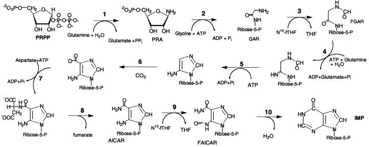
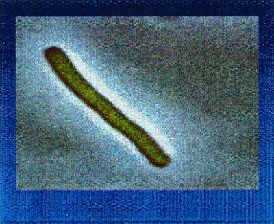
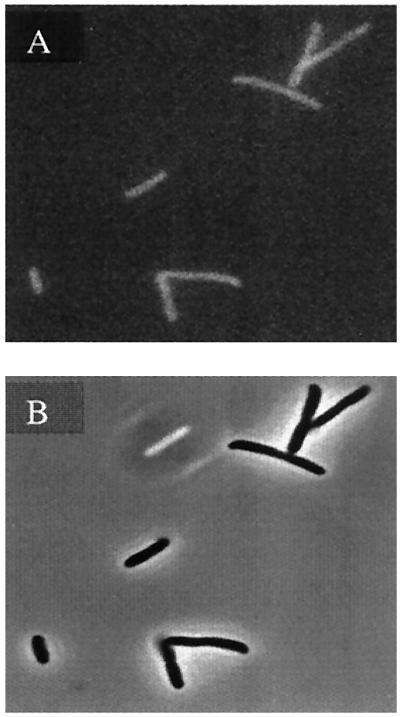
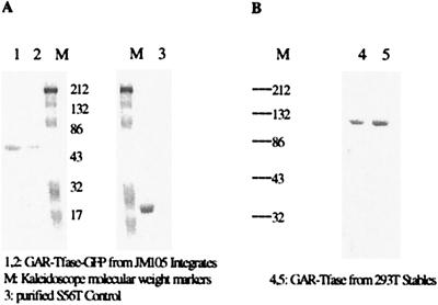
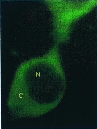
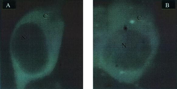
Enzymes of the de novo purine biosynthetic pathway may form a multienzyme complex to facilitate substrate flux through the ten serial steps constituting the pathway. One likely strategy for complex formation is the use of a structural scaffold such as the cytoskeletal network or subcellular membrane of the cell to mediate protein-protein interactions. To ascertain whether this strategy pertains to the de novo purine enzymes, the localization pattern of the third purine enzyme, glycinamide ribonucleotide transformylase (GAR Tfase) was monitored in live Escherichia coli and mammalian cells. Genes encoding human as well as E. coli GAR Tfase fused with green fluorescent protein (GFP) were introduced into their respective cells with regulated expression of proteins and localization patterns monitored by using confocal fluorescence microscopy. In both instances images showed proteins to be diffused throughout the cytoplasm. Thus, GAR Tfase is not localized to an existing cellular architecture, so this device is probably not used to concentrate the members of the pathway. However, discrete clusters of the pathway may still exist throughout the cytoplasm.


Figures

Figure 1
Figure 1
The de novo purine biosynthetic pathway. It is a ten-step pathway involving the conversion of phosphoribosylpyrophosphate to inosine monophosphate. All substrates and intermediates in the pathway are labeled. The third step is catalyzed by the enzyme glycinamide ribonucleotide transformylase (GAR Tfase). This enzyme is monofunctional in E. coli, but exists as part of a trifunctional protein in mammalian cells. The three activities that are condensed are GAR Tfase, GAR synthase, and AIR synthase. The latter enzymes catalyze step 2 and step 5 of purine biosynthesis. Aminoimidazole ribonucleotide transformylase (AICAR Tfase) catalyzes step 9 and exists as a bifunctional, condensed with the tenth activity of the pathway inosine monophosphate (IMP) cyclohydrolase from E. coli to humans. AICAR Tfase uses the same 10-formyltetrahydrofolate cofactor as GAR Tfase.
Figure 2
Figure 2
Image of E. coli GAR Tfase-GFP in BL21(DE3). Cells were grown in LB at 25°C, induced with 50 μM IPTG, and imaged with an Olympus BX 60 microscope equipped with a Micromax cooled charge-coupled device camera driven by the METAMORPH software package. Images were captured by using a U-MWIB excitation cube (Olympus) with a band-pass excitation filter (460–490 nm) and a long pass barrier filter (≥515 nm).
Figure 3
Figure 3
Image of GAR Tfase-GFP Integration Strains. (A) GFP fluorescence image. (B) Phase contrast image of the JM105 integrate strains.
Figure 4
Figure 4
(A) Western analysis of JM105 integrates. (B) Western analysis of the 293 T stably transfected cells. Westerns were performed by using primary rabbit polyclonal anti-GFP antibodies (Molecular Probes) and secondary goat anti-rabbit alkaline phosphatase linked secondary antibody, and developed with nitroblue tetrazolium (NBT) and BCIP (5-bromo-4-chloro-3-indoyl phosphate). Lane 1 and lane 2 are GAR Tfase-GFP from JM105 integrates and lane 3 is a control using purified S65T GFP. (B) Western analysis from 293 T stably transfected cells. M, kaleidoscope molecular weight markers.
Figure 5
Figure 5
Image of Cos-7 cells transiently transfected with pTGFP. Cells were imaged 48 h after transfection. The cells were imaged by using an axiovert microscope attached to a LSM 410 confocal microscope (Zeiss), using 488 nm (argon laser), 488 dichroic, and 515–560 nm emission filtration. Under these imaging conditions, the low level of punctate autofluorescence represented <10% of the GFP signal. The nucleus and cytoplasm are labeled N and C, respectively.
Figure 6
Figure 6
Stable trifunctional GAR Tfase-GFP transfectants of 293 T cells. (A) 293 T cells expressing the trifunctional GAR Tfase-GFP. (B) 293 T fibroblasts expressing GFP. Cells were grown on coverslips, washed twice with 1× PBS, and imaged. The nucleus and cytoplasm are labeled N and C, respectively.

References

    1. Warren M S, Mattia K M, Marolewski A, Benkovic S J. Pure Appl Chem. 1996;68:2029–2036.
    1. Elion G B. Science. 1989;244:41. - PubMed
    1. Seeger D R, Cosulich D B, Smith J R, Hultquist M E. J Am Chem Soc. 1949;71:1753–1758.
    1. Inglese J, Johnson D L, Shiau A, Smith J M, Benkovic S J. Biochemistry. 1990;29:1436–1443. - PubMed
    1. Shim J H, Benkovic S J. Biochemistry. 1998;37:8776–8782. - PubMed

Publication types

Substances

LinkOut - more resources