. 2001 Jun;117(6):573-82.
doi: 10.1085/jgp.117.6.573.

Role of aquaporin water channels in airway fluid transport, humidification, and surface liquid hydration

Affiliations

Role of aquaporin water channels in airway fluid transport, humidification, and surface liquid hydration

Y Song et al. J Gen Physiol. 2001 Jun.

Abstract

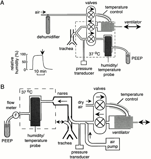
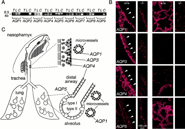
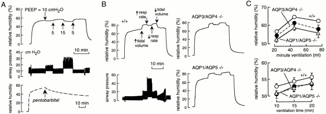
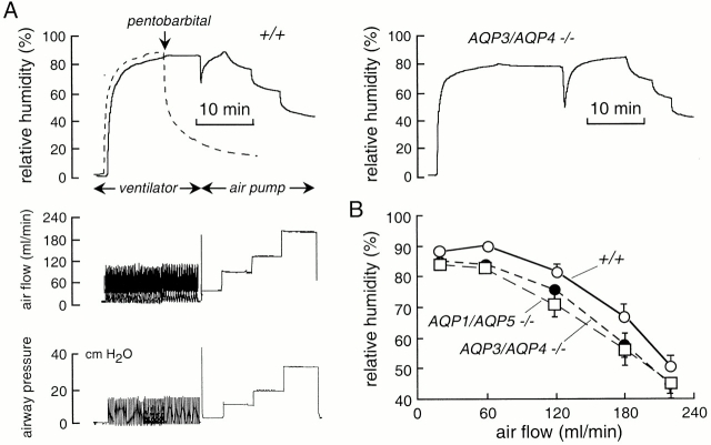
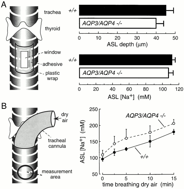
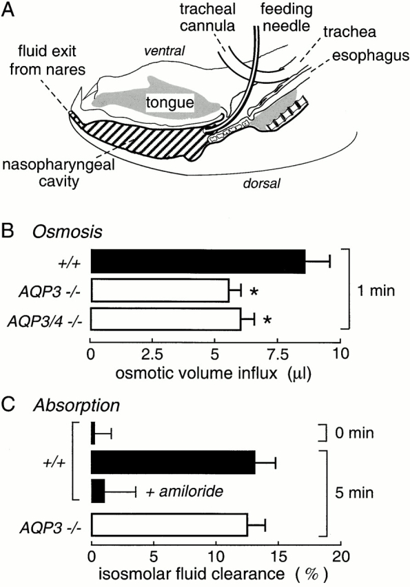
Several aquaporin-type water channels are expressed in mammalian airways and lung: AQP1 in microvascular endothelia, AQP3 in upper airway epithelia, AQP4 in upper and lower airway epithelia, and AQP5 in alveolar epithelia. Novel quantitative methods were developed to compare airway fluid transport-related functions in wild-type mice and knockout mice deficient in these aquaporins. Lower airway humidification, measured from the moisture content of expired air during mechanical ventilation with dry air through a tracheotomy, was 54-56% efficient in wild-type mice, and reduced by only 3-4% in AQP1/AQP5 or AQP3/AQP4 double knockout mice. Upper airway humidification, measured from the moisture gained by dry air passed through the upper airways in mice breathing through a tracheotomy, decreased from 91 to 50% with increasing ventilation from 20 to 220 ml/min, and reduced by 3-5% in AQP3/AQP4 knockout mice. The depth and salt concentration of the airway surface liquid in trachea was measured in vivo using fluorescent probes and confocal and ratio imaging microscopy. Airway surface liquid depth was 45 +/- 5 microm and [Na(+)] was 115 +/- 4 mM in wild-type mice, and not significantly different in AQP3/AQP4 knockout mice. Osmotic water permeability in upper airways, measured by an in vivo instillation/sample method, was reduced by approximately 40% by AQP3/AQP4 deletion. In doing these measurements, we discovered a novel amiloride-sensitive isosmolar fluid absorption process in upper airways (13% in 5 min) that was not affected by aquaporin deletion. These results establish the fluid transporting properties of mouse airways, and indicate that aquaporins play at most a minor role in airway humidification, ASL hydration, and isosmolar fluid absorption.


Figures

Figure 1
Figure 1
Instrumentation for measurement of airway humidification in mice. (A) Measurement of lower airway humidification. After anesthesia, the mouse trachea is cannulated for mechanical ventilation with dry air. Expired air is directed into a chamber containing a humidity sensor. (inset) System efficiency measured using an artificial lung system delivering 100% humidified air. See results for details. (B) Measurement of upper airway humidification. After anesthesia, a cannula is inserted in the low trachea to permit spontaneous breathing. Dry air from a ventilator or air pump (for rapid constant flow) is passed into a second cannula inserted in the proximal trachea. Air exiting the nares passes into a chamber containing the humidity probe.
Figure 2
Figure 2
Aquaporin water channel expression in mouse airways and lung. (A) RT-PCR analysis of aquaporin transcript expression in trachea (T) and peripheral lung (L). Transcripts corresponding to ∼0.3-kb coding sequence fragments of each mouse aquaporin were PCR-amplified using specific primers. Lanes labeled C correspond to amplifications done using a mixture of cDNAs from brain, lung, liver, and kidney, which contained all mouse aquaporins. (B) Immunofluorescence localization of aquaporins 1, 3, 4, and 5 in trachea (wild-type mice, column 1; corresponding knockout mice, column 2) and lung (wild-type mice, column 3, knockout mice, column 4). (arrowheads) Luminal membrane of tracheal epithelium. (C) Schematic of aquaporin expression in airways and lung.
Figure 3
Figure 3
Role of aquaporins in lower airway humidification. (A) Time course of expired air humidity (top) and airway pressure (middle) in response to indicated PEEPs. Tidal volume was 8 ml/kg body weight, and the ventilatory rate was 100/min. Bottom curve shows expired air humidity after pentobarbital overdose and cessation of circulation by transection of the abdominal aorta. (B) Representative time courses of expired air humidity in wild-type and double knockout mice of indicated genotype. Also shown (left, bottom) is corresponding airway pressure for wild-type mouse. Initially, mice were ventilated at 100 breaths/min with 8 ml/kg tidal volume. Where indicated, tidal volume was increased to 15 ml/kg and ventilatory rate to 160 per minute. (C) Summary of experiments as done in B (mean ± SEM, n = 4–5 mice) showing expired air humidity at different minute ventilation (top) and at indicated times after ventilation with dry air (bottom).
Figure 4
Figure 4
Role of aquaporins in upper airway humidification. (A, left) Representative time course of humidity (top), air flow (middle), and airway pressure (bottom) in a wild-type mouse. (right) Same study done on an AQP3/AQP4 double knockout mouse. (B) Averaged relative humidity (mean ± SEM, n = 5 mice) in wild-type mice, and AQP3/AQP4 and AQP1/AQP5 double knockout mice as a function of minute ventilation.
Figure 5
Figure 5
Role of aquaporins in hydration of the airway surface liquid (ASL). (A, left) Schematic of approach to measure ASL depth and [Na+] in anesthetized mice in which a window was created in the trachea for fluorescent dye instillation and z-scanning fluorescence confocal microscopy. (right) ASL depth and [Na+] in wild-type and AQP3/AQP4 double knockout mice (mean ± SEM, n = 4 mice). (B) Measurement of ASL [Na+] during dry air breathing. (left) Fluorescence was measured through the translucent tracheal wall (“measurement area”) below the tip of a tracheal cannula. (right) Time course of ASL [Na+] in wild-type and AQP3/AQP4 double knockout mice (mean ± SEM, n = 4 mice) in response to dry air breathing. Differences in A and B are not significant.
Figure 6
Figure 6
Osmotically driven water transport and isosmolar fluid absorption in upper airways. (A) Sagittal section of mouse head showing air spaces in the nasopharnyx and trachea. The trachea was cannulated to permit spontaneous breathing, and isosmolar or hyperosmolar fluid was instilled into the nasopharyngeal cavity by a feeding needle. Fluid was collected into preweighed vials at specified times by passing air through the feeding needle to expel fluid through the nares. The instilled fluid contained 131I-albumin as a volume marker. Data shown as mean ± SEM, with 6–8 mice per condition. See results for details. (B) Osmotically driven volume influx measured from dilution of the 131I-albumin marker after instillation of 50 μl of a hyperosmolar solution (500 mOsm). (C) Isosmolar fluid absorption (clearance) measured from the increased 131I-albumin concentration at 5 min after instillation of 50 μl of an isosmolar solution (0 min control shown also). Where indicated, 1 mM amiloride was present in the instillate. See results for explanations. *P < 0.01.

Similar articles

Cited by

References

    1. Anderson S.D., Schoeffel R.E., Follet R., Perry C.P., Daviskas E., Kendall M. Sensitivity to heat and water loss at rest and during exercise in asthma patients. Eur. J. Respir. Dis. 1982;63:459–471. - PubMed
    1. Bai C., Fukuda N., Song Y., Ma T., Matthay M.A., Verkman A.S. Lung fluid transport in aquaporin-1 and aquaporin-4 knockout mice. J. Clin. Invest. 1999;103:555–561. - PMC - PubMed
    1. Boucher R.C. Molecular insights into the physiology of the ‘thin film’ of airway surface liquid. J. Physiol. 1999;516:631–638. - PMC - PubMed
    1. Carter E.P., Matthay M.A., Farinas J., Verkman A.S. Transalveolar osmotic and diffusional water permeability in intact mouse lung measured by a novel surface fluorescence method. J. Gen. Physiol. 1996;108:133–142. - PMC - PubMed
    1. Daviskas E., Gonda I., Anderson S.D. Mathematical modeling of heat and water transport in human respiratory tract. J. Appl. Physiol. 1990;69:362–372. - PubMed

Publication types