. 2001 Jun 1;20(11):2757-67.
doi: 10.1093/emboj/20.11.2757.

Activated MEK5 induces serial assembly of sarcomeres and eccentric cardiac hypertrophy

Affiliations

Activated MEK5 induces serial assembly of sarcomeres and eccentric cardiac hypertrophy

R L Nicol et al. EMBO J. .

Abstract

Mitogen-activated protein kinase (MAPK) pathways couple intrinsic and extrinsic signals to hypertrophic growth of cardiomyocytes. The MAPK kinase MEK5 activates the MAPK ERK5. To investigate the potential involvement of MEK5-ERK5 in cardiac hypertrophy, we expressed constitutively active and dominant-negative forms of MEK5 in cardiomyocytes in vitro. MEK5 induced a form of hypertrophy in which cardiomyocytes acquired an elongated morphology and sarcomeres were assembled in a serial manner. The cytokine leukemia inhibitory factor (LIF), which stimulates MEK5 activity, evoked a similar response. Moreover, a dominant-negative MEK5 mutant specifically blocked LIF-induced elongation of cardiomyocytes and reduced expression of fetal cardiac genes without blocking other aspects of LIF-induced hypertrophy. Consistent with the ability of MEK5 to induce serial assembly of sarcomeres in vitro, cardiac-specific expression of activated MEK5 in transgenic mice resulted in eccentric cardiac hypertrophy that progressed to dilated cardiomyopathy and sudden death. These findings reveal a specific role for MEK5-ERK5 in the induction of eccentric cardiac hypertrophy and in transduction of cytokine signals that regulate serial sarcomere assembly.


Figures
None
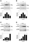
Fig. 1. Activation of endogenous ERK5 by hypertrophic and stress agents. Serum-deprived neonatal rat cardiomyoctyes were treated with (A) 100 µM PE, (B) 1000 U/ml LIF, (C) 200 µM H2O2 and (D) 0.3 M sorbitol for the indicated times, harvested and ERK5 kinase activity was measured. Top: ERK5 was immunoprecipitated from 200 µg of cellular lysate with an antibody specific for the C-terminal 20 amino acids. Kinase assays were performed with immunoprecipitated ERK5 using GST–MEF2C substrate in the presence of [γ-32P]ATP. GST–MEF2C phosphorylation was detected by autoradiography after SDS–PAGE. Middle: immunoblotting was performed on immuno precipitated material using rabbit anti-ERK5 antibody. Bottom: levels of 32P-phosphorylated GST–MEF2C were quantitated with a PhosphorImager. The averaged result ± SD of three independent experiments is shown.
None
Fig. 2. Activated MEK5 induces elongation of cultured neonatal rat cardiomyocytes. Adenoviruses expressing HA-tagged MEK5KM, MEK5WT and MEK5DD were used to infect COS cells at an m.o.i. of 100. (A) Lysates were prepared 48 h post-infection and 5 µg of protein were separated by SDS–PAGE and immunoblotted with anti-HA antibody. (B) Immunoprecipitations were performed on 100 µg of protein with anti-HA antibody. Kinase assays were performed with immunoprecipitated HA-MEK5 using GST–ERK5KMΔ substrate in the presence of [γ-32P]ATP. GST–ERK5KMΔ phosphorylation was detected after SDS–PAGE by autoradiography. Serum-deprived cardiomyocytes were infected at an m.o.i. of 100 with adenovirus expressing (C) β-galactosidase, (D) MEK1CA and (E and G) MEK5DD, or not infected and treated with (F and H) PE (100 µM). Cells were fixed 72 h post-infection and immunostained with anti-sarcomeric α-actinin antibody. Note that cells in (G) and (H) are shown at higher magnification than cells in (C–F). Bar, 20 µm.
None
Fig. 3. Dominant-negative MEK5 blocks LIF-induced elongation of neonatal rat cardiomyocytes. Cardiomyocytes were either not infected or infected with adenovirus at an m.o.i. of 100, serum deprived, and at 24 h post-infection treated either with LIF (1000 U/ml) or PE (100 µM) for an additional 48 h prior to fixation and immunostaining with anti-sarcomeric α-actinin. (A) Uninfected cells treated with LIF. (B) AdMEK5KM-infected cells treated with LIF. (C) Adβ-gal-infected cells treated with LIF. (D) Uninfected cells treated with PE. (E) AdMEK5KM-infected cells treated with PE. (F) Adβ-gal-infected cells treated with PE. Bar, 20 µm.
None
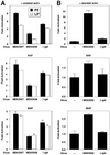
Fig. 4. MEK5 signaling contributes to the regulation of cardiomyocyte fetal gene expression by PE and LIF. (A) Cardiomyocytes were either not infected or infected with MEK5WT, MEK5KM or β-gal adenoviruses at an m.o.i. of 20 and serum deprived. At 36 h post-infection, cells were either not treated or treated with 50 µM PE (black bar) or 1000 U/ml LIF (white bar) for an additional 24 h. RNA was prepared and used for dot-blots with oligonucleotide probes specific for skeletal α-actin, ANF or BNP. Signal intensity was quantitated using a PhosphorImager. The average fold induction ± SD of three independent experiments is shown. Fold induction is relative to uninfected cells without PE or LIF treatment. (B) Cardiomyocytes were either not infected (–) or infected with MEK5DD or β-gal adenoviruses at an m.o.i. of 20 and serum deprived. At 48 h post-infection, the cells were harvested and RNA was prepared. Transcript levels for α-skeletal actin, ANF or BNP were determined as described in (A). Fold induction is relative to uninfected cells.
None
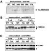
Fig. 5. Expression of MEK5 and ERK5 in wild-type and MEK5DD transgenic mice. Lysates were prepared from wild-type (WT) and transgenic (TG) hearts, and 20 µg of protein were separated by SDS–PAGE. (A) Expression of HA-tagged MEK5DD was analyzed in different lines of transgenic mice by immunoblotting with anti-HA antibody. Lines of MEK5DD transgenic mice are indicated by identifying numbers. For each line, lysate was prepared from two hearts and loaded in adjacent lanes. Expression of (B) MEK5 and (C) ERK5 was analyzed in wild-type and line 367 MEK5DD transgenic mice by immunoblotting with L610 rabbit anti-MEK5 antiserum and rabbit anti-ERK5. Bands that are either non-specific (asterisk) or degradation products (arrowhead) are indicated. Note the reduced mobility of ERK5 in transgenic animals relative to wild type.
None
Fig. 6. Survival curve for wild-type and MEK5DD transgenic mice. F1 hemizygous transgenic mice were generated by backcrossing the transgenic founder mouse with C57B6 mice. The open circles represent the percentage survival of wild-type (WT) F1 mice (n = 24); the closed circles represent percentage survival of transgenic (TG) F1 mice (n = 24).
None
Fig. 7. MEK5DD transgenic hearts show progressive dilation and thinning of ventricular walls with age. (A) Hearts were removed from wild-type and MEK5DD transgenic mice at 3, 6 and 12 weeks of age. Hearts were fixed in 10% PBS-buffered formalin and photographed. (B) Hearts from 12-week-old MEK5DD-transgenic and wild-type mice were fixed and sectioned longitudinally or at the midsagittal level parallel to the base and stained with hematoxylin–eosin. ra, right atrium; la, left atrium; rv, right ventricle; lv, left ventricle.
None
Fig. 8. MEK5DD transgenic hearts show reduced myofiber cross-sectional area relative to wild type. Hearts were removed from 8-week-old wild-type and from MEK5DD and calcineurin transgenic mice, fixed, sectioned and stained with hematoxylin–eosin. Dramatic differences in myocyte cross-sectional area are apparent in hematoxylin–eosin-stained sections from (A) MEK5DD transgenic hearts, (B) wild-type hearts and (C) calcineurin transgenic hearts. Bar, 20 µm. (D) The cross-sectional area of myocytes from 8-week-old wild-type and MEK5DD transgenic mice was quantitated using a computerized morphometric system. Measurements were made on equivalent sections from five wild-type and five transgenic hearts, and, within each section, measurements were taken from left and right ventricle, septum and papillary muscle (10 measurements each). The average result ± SD is shown; *P <0.001.
None
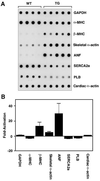
Fig. 9. Induction of fetal gene expression in MEK5DD transgenic hearts. RNA was prepared from wild-type and transgenic hearts. (A) RNA dot-blots were prepared with 1 µg of RNA/dot and probed with oligonucleotide probes specific for the indicated gene. (B) The average fold induction or repression of gene expression ± SD for MEK5DD transgenic animals relative to wild type is shown. Signal intensity was quantitated using a PhosphorImager.
None
Fig. 10. A model for LIF-induced cardiomyocyte elongation mediated by MEK5 inhibition of parallel assembly of sarcomeres. MEK5 induces cardiomyocyte elongation by interfering with parallel assembly of sarcomeres. Other signaling molecules implicated downstream of LIF include MEK1, JAK/STAT, PI3-K, CaMK and calcineurin.

Similar articles

Cited by

References

    1. Abe J., Kusuhara,M., Ulevitch,R.J., Berk,B.C. and Lee,J.D. (1996) Big mitogen-activated protein kinase 1 (BMK1) is a redox-sensitive kinase. J. Biol. Chem., 271, 16586–16590. - PubMed
    1. Abe J., Baines,C.P. and Berk,B.C. (2000) Role of mitogen-activated protein kinases in ischemia and reperfusion injury: the good and the bad. Circ. Res., 86, 607–609. - PubMed
    1. Adams J.W., Sakata,Y., Davis,M.G., Sah,V.P., Wang,Y., Liggett,S.B., Chien,K.R., Brown,J.H. and Dorn,G.W. (1998) Enhanced Gαq signaling: a common pathway mediates cardiac hypertrophy and apoptotic heart failure. Proc. Natl Acad. Sci. USA, 95, 10140–10145. - PMC - PubMed
    1. Becker T.C., Noel,R.J., Coats,W.S., Gomez-Foix,A.M., Alam,T., Gerard,R.D. and Newgard,C.B. (1994) Use of recombinant adenovirus for metabolic engineering of mammalian cells. Methods Cell Biol., 43, 161–189. - PubMed
    1. Bueno O.F. et al. (2000) The MEK1–ERK1/2 signaling pathway promotes compensated cardiac hypertrophy in transgenic mice. EMBO J., 19, 6341–6350. - PMC - PubMed

Publication types

MeSH terms