. 2001 Jun 1;20(11):2802-11.
doi: 10.1093/emboj/20.11.2802.

Hormone-induced nucleosome positioning in the MMTV promoter is reversible

Affiliations

Hormone-induced nucleosome positioning in the MMTV promoter is reversible

S Belikov et al. EMBO J. .

Abstract

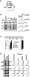
The mouse mammary tumor virus (MMTV) promoter is induced by glucocorticoid hormone via the glucocorticoid receptor (GR). The hormone-triggered effects on MMTV transcription and chromatin structure were studied in Xenopus oocytes. We previously showed that the nucleosomes organizing the MMTV promoter became translationally positioned upon hormone induction. A single GR-binding site was necessary and sufficient for the chromatin events to occur, while transcription and basal promoter elements were dispensable. Here we show that addition of the hormone antagonists RU486 or RU43044 to the previously hormone-induced MMTV promoter results in cessation of transcription and loss of chromatin remodeling and nucleosome positioning. In vivo footprinting demonstrated agonist- and RU486-induced GR binding to its DNA response element (GRE), while the other antagonist, RU43044, did not promote GR-GRE interaction. These results demonstrate that induction and maintenance of nucleosome positioning is an active process that requires constant 'pressure' of agonist-GR-recruited chromatin-modifying factor(s) rather than GR-DNA binding itself.


Figures

None
Fig. 1. The reporter DNA construct, the pMTV:M13 used for injection with the primers used for primer extension analysis of the SacI in situ accessibility assay and DMS methylation protection (solid black arrows), and the restriction enzyme cleavage sites that are referred to in the text. White boxes designate GRE hexanucleotide elements I–IV; the black box (Truss et al., 1995) designates an NF1 site; light gray boxes designate OCT1 sites; and the dark gray box designates the TATA sequence. The B-nucleosome probe used in MNase experiments is shown below.
None
Fig. 2. RU43044 repressed agonist-driven MMTV transcription and reversed chromatin remodeling and nucleosome positioning. (A) Schematic representation of the experimental design. A 100 nM concentration of Cort and 50 µM RU43044 (RU044) were added at the indicated times. A = control; B = agonist; C = antagonist added after agonist; D = agonist + antagonist added at the same time. The 5 and 10 designate hours ± hormone incubation for A and B incubations. (B) Transcription analysis by S1 nuclease protection of MMTV and AdML RNA. Effect of addition of RU43044 on MMTV transcription analyzed in whole oocytes (lanes 1–5) and in manually isolated nuclei (lanes 6–8), six oocytes per group. The diagram below shows quantification of radioactivity. One oocyte equivalent per lane was present in lanes 1–5 and six nuclei per lane in 6–8. B10 is set to 100%. (CSacI accessibility assay, six oocytes per group. Arrows show specific bands generated by SacI and HinfI as developed by primer extension. DNA shows the analysis with a naked DNA control. The staple diagram below shows the quantification; SacI cutting is given as a percentage of total DNA. (D) Effect of addition of RU43044 on DNA topology of MMTV minichromosomes extracted from the Xenopus oocytes. Radioactivity scans of the lanes are shown below. (E) Effect of addition of RU43044 on nucleosomal array organization. Groups of 10 oocytes were injected and treated with hormone agonist/antagonist as indicated. Oocytes were harvested and digested with increasing amounts of MNase. DNA was resolved in an agarose gel, transferred and hybridized with a labeled MMTV promoter probe encompassing region –192/–100 (B-nucleosome probe) and then washed and rehybridized with an M13 vector probe (below). The black dot shows the subnucleosomal DNA fragment of ∼120 bp revealed only after hybridization with the specific probe. Radioactivity scans of lanes with corresponding maximum MNase concentration are shown to the right. (F) Effect of addition of RU43044 on nucleosome positioning over the MMTV promoter. Groups of seven oocytes, injected and treated as indicated, were digested with MPE for 3 and 10 min. Isolated DNA was digested with EcoRV (+425), resolved on an agarose gel, blotted and hybridized with a random-primed labeled fragment adjacent to the EcoRV site (EcoRV–SacI fragment). M, internal molecular weight markers; DNA, naked dsMMTV DNA digested with MPE. To the right is a schematic summary of MPE cuts with putative nucleosome positions. Radioactivity scans of corresponding lanes are shown to the right.
None
Fig. 3. RU486 represses agonist-driven MMTV transcription and reverses chromatin remodeling and nucleosome positioning. (AF) As described in Figure 2 legend.
None
Fig. 4. Increasing hormone (TA) concentration induces GR nuclear translocation, MMTV transcription, chromatin remodeling and nucleosome positioning. (A) Nuclear translocation of GR. Oocytes were injected with GR mRNA and pMTV:M13 ssDNA. On the next day, hormone (TA) at the indicated concentrations (nM) was added; after 8 h, oocytes were harvested, manually dissected in groups of five and GR content was evaluated separately in nucleus and cytoplasm by immunoblotting. Relative cytosol/nuclear amounts are given below. (B) Effect of hormone concentration on MMTV transcription assayed by S1 nuclease protection. Six oocytes per group were homogenized and analyzed separately for each TA concentration. (C) Effect of hormone concentration on SacI accessibility assay of MMTV DNA. Six oocytes per group were analyzed for each hormone concentration. See legend of Figure 2C for details. (D) Effect of hormone concentration on nucleosomal array organization in the MMTV promoter. Groups of 10 oocytes were MNase digested and DNA was processed as indicated in Figure 2E. (E) Effect of hormone concentration on nucleosome positioning over the MMTV promoter. Groups of seven oocytes were homogenized and digested with MPE and analyzed as indicted in Figure 2F. M, internal molecular weight markers; DNA, naked MMTV dsDNA digested with MPE. To the right is a schematic summary of MPE cuts with putative nucleosome positions. Radioactivity scans are shown to the right.
None
Fig. 5. Agonist- and antagonist-induced nuclear uptake and GR–GRE binding. (A) Nuclear translocation of hormone/antagonists-liganded GR. Oocytes were injected with GR mRNA and the next day treated with agonist (1 µM TA) or antagonists (50 µM RU486 or RU43044), respectively. Eight hours later, oocytes, five per group, were harvested and GR content in manually dissected nuclei was evaluated by immunoblotting. Mock designates non-injected oocytes. (B) DMS methylation protection over the GRE I segment. Oocytes in groups of five were treated with DMS (see Materials and methods). The methylation pattern was developed by primer (–291/–265) extension. Corresponding Gs are indicated with arrows (white arrows indicate protected guanosines). Radioactivity scans of lanes corresponding to the maximum DMS concentration are shown to the right. (C) The sequence of the GRE I segment with the boxed GR-binding sites and the mean value of the quantification of two separate DMS protection experiments. The –165 G was used as internal standard. (D) DMS methylation protection over the GRE II–IV segments. The methylation pattern was developed by primer (+92/+65) extension. Protected Gs within corresponding GREs are indicated with white arrows. Radioactivity scans of lanes are shown to the right.

Similar articles

Cited by

References

    1. Agalioti T., Lomvardas,S., Parekh,B., Yie,J., Maniatis,T., Thanos,D., El-Osta,A. and Wolffe,A.P. (2000) Ordered recruitment of chromatin modifying and general transcription factors to the IFN-β promoter. Cell, 103, 667–678. - PubMed
    1. Almouzni G. and Wolffe,A.P. (1993) Replication-coupled chromatin assembly is required for repression of basal transcription in vivo. Genes Dev., 7, 2033–2047. - PubMed
    1. Archer T.K., Cordingley,M.G., Wolford,R.G. and Hager,G.L. (1991) Transcription factor access is mediated by accurately positioned nucleosomes on the mouse mammary tumor virus promoter. Mol. Cell. Biol., 11, 688–698. - PMC - PubMed
    1. Beato M. and Eisfeld,K. (1997) Transcription factor access to chromatin. Nucleic Acids Res., 25, 3559–3563. - PMC - PubMed
    1. Becker P.B., Gloss,B., Schmid,W., Strahle,U. and Schutz,G. (1986) In vivo protein–DNA interactions in a glucocorticoid response element require the presence of the hormone. Nature, 324, 686–688. - PubMed

Publication types

MeSH terms