

Epidermal growth factor receptor-targeted immunophotodiagnosis and photoimmunotherapy of oral precancer in vivo
- PMID: 11389080
- PMCID: PMC3441050
Epidermal growth factor receptor-targeted immunophotodiagnosis and photoimmunotherapy of oral precancer in vivo
Abstract
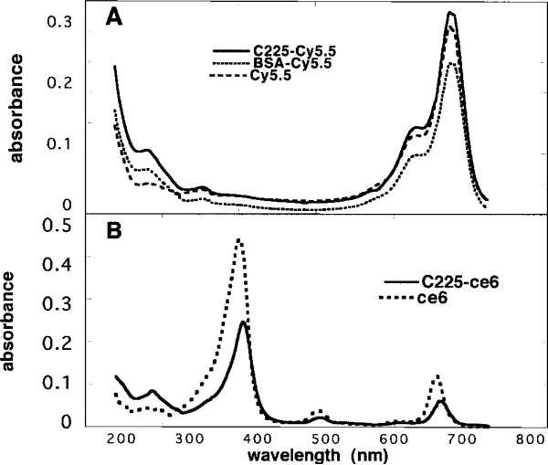
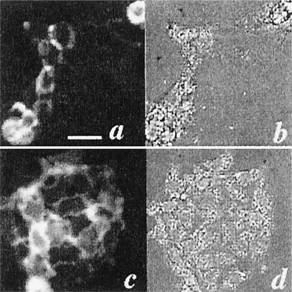
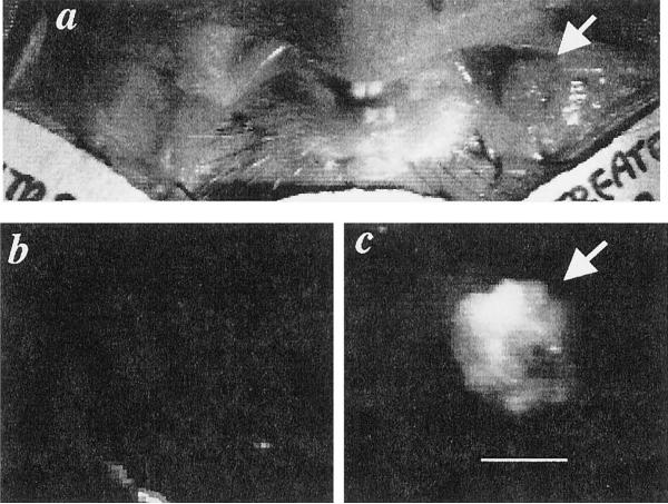
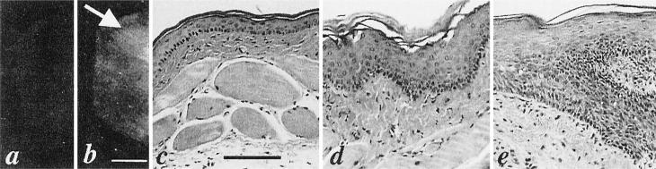
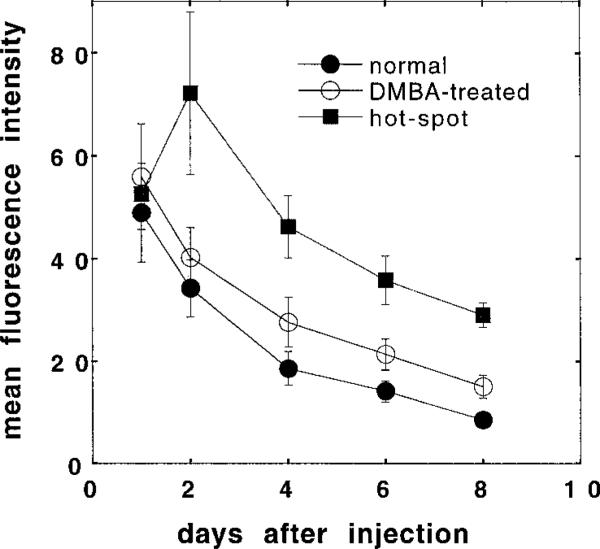
Immunophotodiagnosis uses a fluorescence-labeled monoclonal antibody (MAb) that recognizes a tumor-associated antigen to image the fluorescence emitted from the fluorophore-bound MAb that has localized in the tissue. It may be used to diagnose malignant or precancerous lesions, to delineate the margins for tumor resection, or as a feedback mechanism to assess response to treatment. In oral precancer, the epidermal growth factor receptor (EGFR) is overexpressed and could be used as a marker for early detection or as a target for therapy. The goal of this study was to test an anti-EGFR MAb (C225) coupled to either the near-infrared fluorescent dye N,N'-di-carboxypentyl-indodicarbocyanine-5,5'-disulfonic acid for detection or a photochemically active dye (chlorin(e6)) for therapy of early premalignancy in the hamster cheek pouch carcinogenesis model. Fluorescence levels in the carcinogen-treated tissue correlated with the histological stage of the lesions when the C225-N,N'-di-carboxypentyl-indodicarbocyanine-5,5'-disulfonic acid conjugate was used but did not do so with the irrelevant conjugates. Discrete areas of clinically normal mucosa with high fluorescence (hot spots) were subsequently shown by histology to contain dysplastic areas. The best contrast between normal and carcinogen-treated cheek pouches was found at 4-8 days after injection. To test the potential of immunophotodiagnosis as a feedback modality for therapeutic intervention, experiments were conducted with the same MAb conjugated to chlorin(e6) followed by illumination to reduce expression of the EGFR by a photodynamic effect. Subsequent immunophotodiagnosis showed that this treatment led to a significant reduction in fluorescence in the carcinogen-treated cheek pouch compared with nonilluminated areas. This difference between illuminated and dark areas was not seen in the normal cheek pouch. Taken together, the results demonstrate the potential for development of immunophotodiagnosis as a diagnostic tool and as a method of monitoring response to therapy and that the EGFR may be an appropriate target in head and neck cancer.
Figures
References
-
- Speight PM, Morgan PR. The natural history and pathology of oral cancer and precancer. Community Dent. Health. 1993;10(Suppl. 1):31–41. - PubMed
-
- Scully C. Clinical diagnostic methods for the detection of premalignant and early malignant oral lesions. Community Dent. Health. 1993;10(Suppl. 1):43–52. - PubMed
-
- Eisbruch A, Blick M, Lee JS, Sacks PG, Gutterman J. Analysis of the epidermal growth factor receptor gene in fresh human head and neck tumors. Cancer Res. 1987;47:3603–3605. - PubMed
-
- Shirasuna K, Hayashido Y, Sugiyama M, Yoshioka H, Matsuya T. Immunohistochemical localization of epidermal growth factor (EGF) and EGF receptor in human oral mucosa and its malignancy. Virchows Archiv. A Pathol. Anat. Histol. 1991;418:349–353. - PubMed
-
- Ke LD, Adler-Storthz K, Clayman GL, Yung AW, Chen Z. Differential expression of epidermal growth factor receptor in human head and neck cancers. Head Neck. 1998;20:320–327. - PubMed
Publication types
MeSH terms
Substances
Grants and funding
LinkOut - more resources
Full Text Sources
Other Literature Sources
Medical
Research Materials
Miscellaneous