. 2001 Jun 1;533(Pt 2):571-83.
doi: 10.1111/j.1469-7793.2001.0571a.x.

Eccentric exercise-induced morphological changes in the membrane systems involved in excitation-contraction coupling in rat skeletal muscle

Affiliations

Eccentric exercise-induced morphological changes in the membrane systems involved in excitation-contraction coupling in rat skeletal muscle

H Takekura et al. J Physiol. .

Abstract

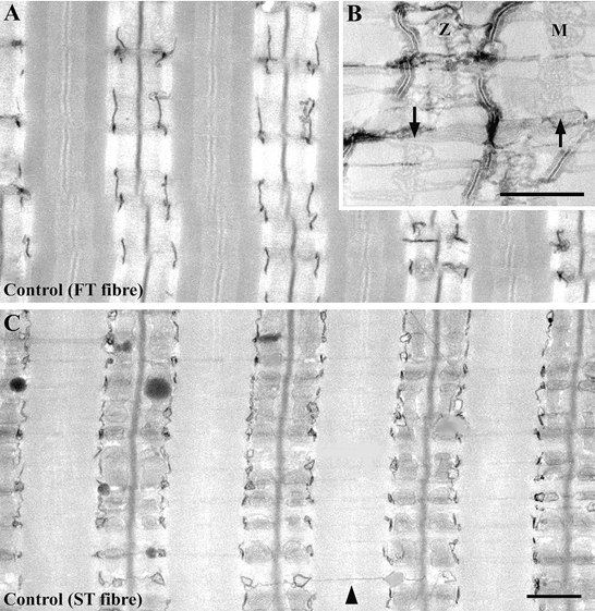
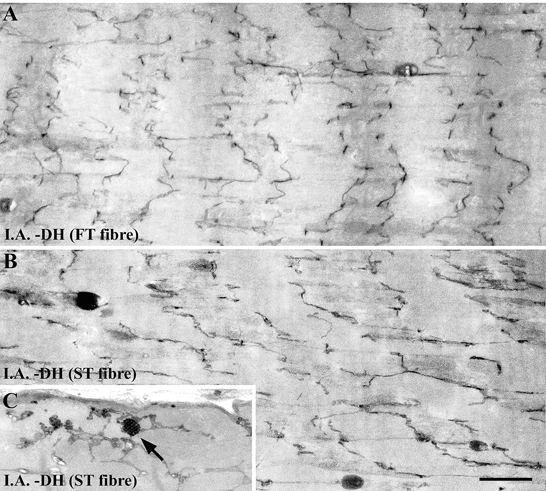
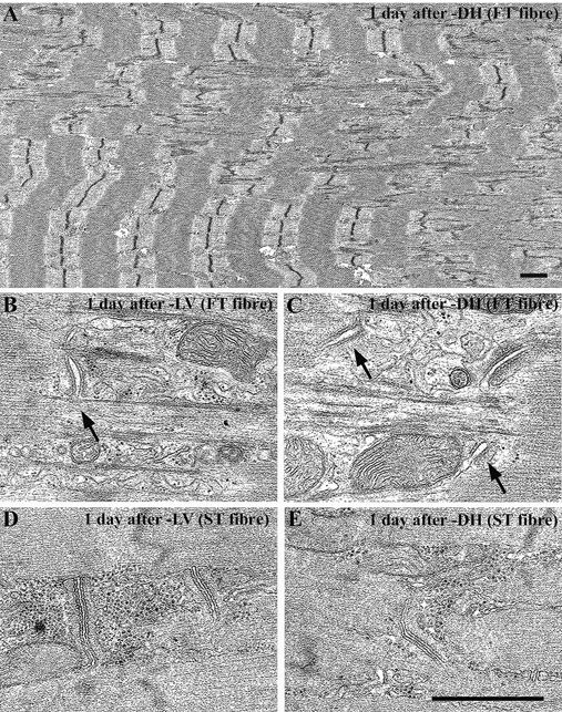
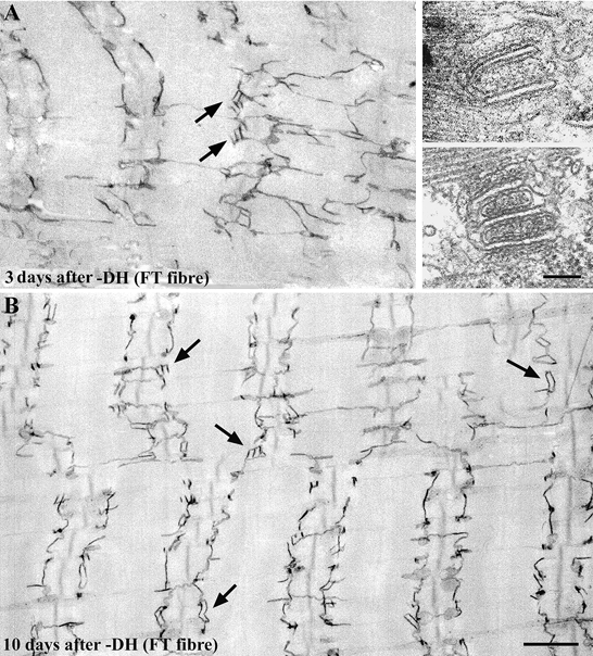
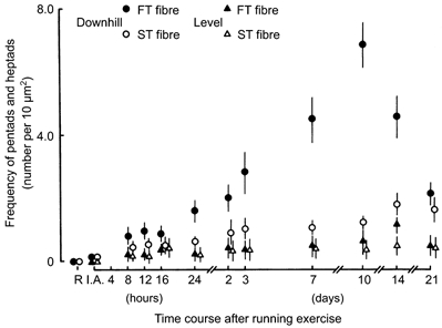
1. Physiological evidence suggests that excitation-contraction (E-C) coupling failure results from eccentric contraction-induced muscle injury because of structural and morphological damage to membrane systems directly associated with the E-C coupling processes within skeletal muscle fibres. In this study using rats, we observed the ultrastructural features of the membrane systems of fast-twitch (FT) and slow-twitch (ST) muscle fibres involved in E-C coupling following level and downhill running exercise. Our aim was to find out whether mechanically mediated events following eccentric exercise caused disorder in the membrane systems involved in E-C coupling, and how soon after exercise such disorder occurred. We also compared the morphological changes of the membrane systems between ST and FT muscle fibres within the same muscles. 2. Single muscle fibres were dissected from triceps brachii muscles of male Fischer 344 rats after level or downhill (16 deg decline) motor-driven treadmill running (18 m min(-1), 5 min running with 2 min rest interval, 18 bouts). All single muscle fibres were histochemically classified into ST or FT fibres. The membrane systems were visualized using Ca(2+)-K(3)Fe(CN)(6)-OsO(4) techniques, and observed by high voltage electron microscopy (120-200 kV). 3. There were four obvious ultrastructural changes in the arrangement of the transverse (t)-tubules and the disposition of triads after the downhill running exercise: (1) an increase in the number of longitudinal segments of the t-tubule network, (2) changes in the direction and disposition of triads, (3) the appearance of caveolar clusters, and (4) the appearance of pentads and heptads (close apposition of two or three t-tubule elements with three or four elements of terminal cisternae of the sarcoplasmic reticulum). The caveolar clusters appeared almost exclusively in the ST fibres immediately after downhill running exercise and again 16 h later. The pentads and heptads appeared almost exclusively in the FT fibres, and their numbers increased dramatically 2-3 days after the downhill running exercise. 4. The eccentric exercise led to the formation of abnormal membrane systems involved in E-C coupling processes. These systems have unique morphological features, which differ between ST and FT fibres, even within the same skeletal muscle, and the damage appears to be concentrated in the FT fibres. These observations also support the idea that eccentric exercise- induced E-C coupling failure is due to physical and chemical disruption of the membrane systems involved in the E-C coupling process in skeletal muscle.


Figures

Figure 1
Figure 1. Longitudinal sections of fast-twitch (FT; A and B) and slow-twitch (ST; C) fibres in sedentary control rat triceps brachii muscle, following Ca2+-K3Fe(CN)6 treatment
The lumen of the t-tubules is variably filled with electron-dense precipitates, and the precipitate-filled t-tubules appear dark. The SR is more difficult to see from the precipitate and is less intensely stained (B). Note the empty spaces between the t-tubule and SR terminal cisternae, which become apparent at high magnification of the Ca2+-K3Fe(CN)6-treated muscle fibres (B). These empty spaces are the junctional gaps between the two membrane systems. Using an electron microscope, we could observe feet occupying the junctional gap in this section sample. We could not, however, see feet at the junctional gap in the Ca2+-K3Fe(CN)6-treated muscle fibres. The disposition of t-tubules and triads is typically highly ordered in adult skeletal muscle fibres. The SR is well developed not only at the A-I junctional regions that form triads with the t-tubules, but also at the M-line regions (arrows in B; M, M-line; Z, Z-line). It is difficult to see the longitudinally oriented t-tubules across each sarcomere (C, arrow head) in the mature muscle fibres. Scale bars, 1 μm.
Figure 2
Figure 2. Longitudinal sections of FT (A) and ST (B and C) fibres in rat triceps brachii muscle immediately after the downhill running exercise
The focal disruption of the Z-lines demonstrates a different kind of streaming: the Z-lines are wavy and run a zig-zag course. The disruption of the t-tubule network was first observed immediately after (I.A.) the downhill running exercise (DH) in both the FT and ST fibres, shortly after the Z-line disruption appeared. We observed a relatively high number of three-dimensional clusters of caveolae-like structures, which are associated with the t-tubule network in the ST fibre (C, arrow). Scale bar, 1 μm.
Figure 3
Figure 3. Longitudinal thin sections of FT (A-C) and ST (D and E) fibres in rat triceps brachii muscle 1 day after exercise for the level running group (B and D) and for the downhill running group (A, C and E)
Z-line smearing and focal disruption of the A-band region can be seen throughout the FT fibres. The disposition and direction of the triads has changed as a result of the focal loss of the Z-lines, and the central element of the triads and t-tubules is swollen in the FT fibres for both the level running group (B, arrow) and the downhill running group (C, arrows). This structural damage is especially evident in the FT fibres of the downhill running group. Scale bars, 1 μm.
Figure 4
Figure 4. Longitudinal sections of FT fibres in rat triceps brachii muscle at 3 days (A) and 10 days (B) after the exercise for the downhill running group
The peculiar pentadic and heptadic structures (two or three t-tubule elements with three or four elements of terminal cisternae, closely aligned to terminal cisternae of SR, indicated by arrows in A, and shown in the insets), which are seldom found in normal muscle fibres, appeared 1 day after exercise only in the FT fibres. Ultrastructural features of these pentads and heptads, such as the width of the junctional gap and feet, were similar to the features of the triads in the muscle fibres of the control group. The number of pentads and heptads increased dramatically 2-3 days after the exercise. The pentads and heptads are located near the A-I junction region with or without transverse orientation (A, arrows). Interestingly, the number of longitudinal connections of t-tubules seemed to decrease after the number of pentads and heptads increased (B). Scale bars, 1 μm.
Figure 5
Figure 5
Changes in the longitudinally oriented sarcotubular surface area relative to normalized muscle fibre volume in FT and ST fibres in rat triceps brachii muscle following exercise for the level and downhill running groups. The means and standard deviations (mean ± 1 s.d.) are given.
Figure 6
Figure 6
Changes in the frequency of pentads and heptads in FT and ST fibres following exercise in rat triceps brachii muscle for both the level and downhill running groups. The means and standard deviations (mean ± 1 s.d.) are given.

Similar articles

Cited by

References

    1. Appelt D, Buenviaje B, Champ C, Franzini-Armstrong C. Quantitation of ‘junctional feet’ content in two types of muscle fiber from hind limb muscles of the rat. Tissue and Cell. 1989;21:783–794. - PubMed
    1. Armstrong RB, Ogilvie RW, Schwane JA. Eccentric exercise-induced injury to rat skeletal muscle. Journal of Applied Physiology. 1983;54:80–93. - PubMed
    1. Balnave CD, Allen DG. Intracellular calcium and force in single mouse muscle fibres following repeated contraction with stretch. Journal of Physiology. 1995;488:23–36. - PMC - PubMed
    1. Bar PRD, Reijneveld JC, Wokke JHJ, Jacobs SCJM, Bootsma AL. Muscle damage induced by exercise: Nature, prevention and repair. In: Salmons S, editor. Muscle Damage. Oxford, UK: Oxford University Press; 1997. pp. 1–27.
    1. Byrd SK. Alterations in the sarcoplasmic reticulum. A possible link to exercise-induced muscle damage. Medicine and Science in Sports and Exercise. 1992;24:531–536. - PubMed

Publication types

MeSH terms

Substances

LinkOut - more resources