. 2001 Aug 10;276(32):30423-8.
doi: 10.1074/jbc.M101194200. Epub 2001 Jun 4.

Krüppel-like factor 4 (gut-enriched Krüppel-like factor) inhibits cell proliferation by blocking G1/S progression of the cell cycle

Affiliations

Krüppel-like factor 4 (gut-enriched Krüppel-like factor) inhibits cell proliferation by blocking G1/S progression of the cell cycle

X Chen et al. J Biol Chem. .

Abstract

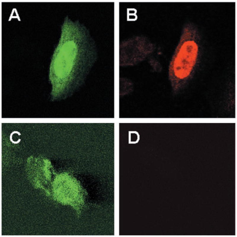
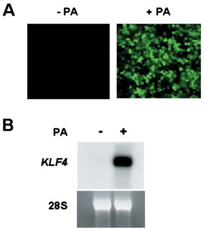
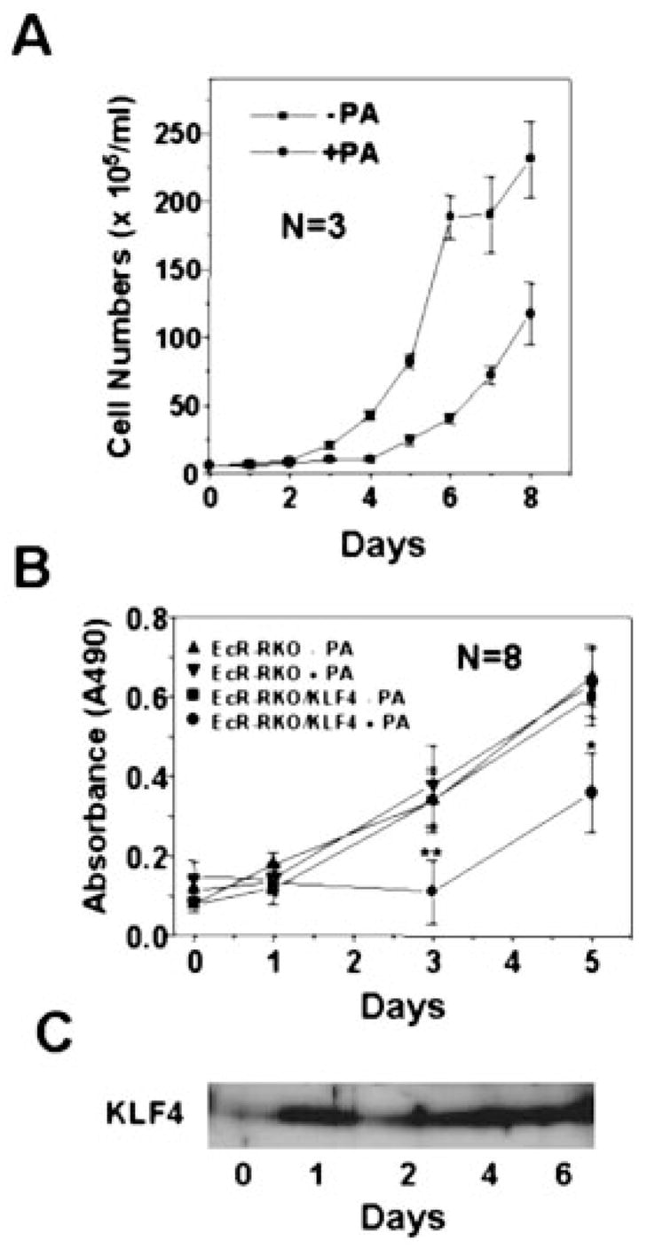
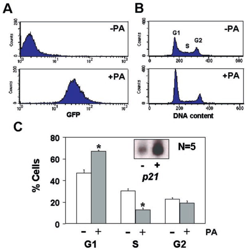
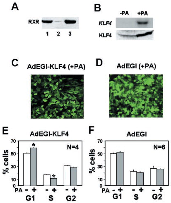
Krüppel-like factor 4 (KLF4) is an epithelial cell-enriched, zinc finger-containing transcription factor, the expression of which is associated with growth arrest. Previous studies show that constitutive expression of KLF4 inhibits DNA synthesis but the manner by which KLF4 exerts this effect is unclear. In the present study, we developed a system in which expression of KLF4 is controlled by a promoter that is induced upon treatment of cells containing the receptors for the insect hormone, ecdysone, with ponasterone A, an ecdysone analogue. The rate of proliferation of a stably transfected colon cancer cell line, RKO, was significantly decreased following addition of ponasterone A when compared with untreated cells. Flow cytometric analyses indicated that the inducible expression of KLF4 caused a block in the G(1)/S phase of the cell cycle. A similar block was observed when ecdysone receptor-containing RKO cells were infected with a replication-defective recombinant adenovirus containing an inducible KLF4 and treated with ponasterone A. Results of these studies provide evidence that the inhibitory effect of KLF4 on cell proliferation is mainly exerted at the G(1)/S boundary of the cell cycle.


Figures

Fig. 1
Fig. 1. Inducible expression of EGFP and KLF4 in transiently transfected cells
EcR-CHO cells were transfected with pAdLoxEGI-KLF4 and treated with 5 μm ponasterone A for 24 h. Cells were fixed and stained with an anti-KLF4 antibody (panel B) or without any primary antibody (panel D), followed by a Cy3-labeled secondary antibody. Shown in panels A and B is a representative cell that contained EGFP (green) and KLF4 (red), respectively. In contrast, neither of the two green cells in panel C stained red in the absence of the primary antibody (panel D) (magnification, ×100).
Fig. 2
Fig. 2. Inducible expression of KLF4 in stably transfected RKO cells
A stable RKO cell line (EcR-RKO/pAdLoxEGI-KLF4) was established by co-transfection with pVgRXR and pAdLoxEGI-KLF4. After treatment with or without 5 μm ponasterone A (PA) for 24 h, cells were inspected under an inverted fluorescence microscope for the presence of GFP (panel A). Panel B shows the result of a Northern blot analysis of RNA isolated from uninduced (−) and induced (+) cells, and probed with a labeled KLF4 cDNA fragment (upper panel). A photograph of the 28 S ribosomal RNA (lower panel) from both sets of cells is included to indicate equal loading.
Fig. 3
Fig. 3. The effect of induced KLF4 expression on proliferation of RKO cells
In panel A, 2.8 × 10⁵ EcR-RKO/pAdLoxEGI-KLF4 cells were seeded and continuously cultured in 60-mm dishes with or without 5 μm ponasterone A for up to 8 days. The media were changed every other day. Cells were counted with a hemocytometer daily. Cell numbers are expressed as mean ± S.D. (bars). In panel B, EcR-RKO and EcR-RKO/pAdLoxEGI-KLF4 (labeled as EcR-RKO/KLF4 in the figure) cells were plated in 96-well microtiter plates at a density of 1 × 10³ cells per well. After 24 h, fresh media containing either 5 μm ponasterone A or vehicle only were added. The number of cells was determined using the colorimetric MTS assay on days 0, 1, 3, and 5. Results are depicted as mean ± S.D of absorbance at 490 nm as a function of time. *, p < 0.01; **, p < 0.0001 by paired Student t test. Panel C is a Western blot analysis of proteins extracted from induced cells for the presence of KLF on the days indicated.
Fig. 4
Fig. 4. The effect of induced KLF4 expression on the cell cycle
EcR-RKO/pAdLoxEGI-KLF4 cells were cultured to ~50% confluency and treated with or without 5 μm ponasterone A for 24 h. Cells were fixed and stained with propidium iodide, and then analyzed by FACS. Panel A shows the result of FACS based on the intensity of green fluorescence in uninduced (−PA) and induced (+PA) cells. Panel B shows the DNA content as revealed by propidium iodide staining in both conditions. DNA contents corresponding to the 3 phases (G1, S, and G2) of the cell cycle are labeled as such. In panel C, the mean percentages of cells with DNA content in each of the 3 phases of the cell cycle under the uninduced or induced condition over five independent determinations were shown as mean ± S.D. (bars). *, p < 0.001. Inset shows the result of a Northern blot analysis for p21WAF1/Cip1 in the absence (−) or presence (+) of ponasterone A.
Fig. 5
Fig. 5. The effect of inducible expression of KLF4 in cells infected with recombinant adenoviruses containing EGFP and KLF4 or EGFP alone
Panel A is a Western blot analysis for RXR production in CHO cells with stably transfected pVgRXR (lane 1), untransfected RKO cells (lane 2), and the stably transfected RKO cells, EcR-RKO (lane 3). In panel B, EcR-RKO cells were infected with the recombinant AdEGI-KLF4 virus and treated (+PA) or not (−PA) with ponasterone A for 24 h and then analyzed for the RNA (upper panel) and protein (lower panel) content for KLF4 by Northern and Western blotting, respectively. In panels C and D, EcR-RKO cells were infected with AdEGI-KLF4 and AdEGI, respectively, and treated with 5 μm ponasterone A for 24 h. Shown are cells observed under a fluorescence microscope. In panels E and F, EcR-RKO cells were infected with AdEGI-KLF4 and AdEGI, respectively, and treated or not with 5 μm ponasterone A for 24 h. Cells were stained with propidium iodide and the DNA content analyzed by FACS. Shown are the mean and S.D. of percentages of cells in the 3 phases of the cell cycle. *, p < 0.001.

References

    1. Shields JM, Christy RJ, Yang VW. J Biol Chem. 1996;271:20009–20017. - PMC - PubMed
    1. Garrett-Sinha LA, Eberspaecher H, Seldin MF, de Crombrugghe B. J Biol Chem. 1996;271:31384–31390. - PubMed
    1. Schuh R, Aicher W, Gual U, Cote S, Preiss A, Maier D, Seifert E, Nauber U, Shroder C, Kemler R, Jackle H. Cell. 1986;47:1025–1032. - PubMed
    1. Miller IJ, Bieker JJ. Mol Cell Biol. 1993;13:2776–2786. - PMC - PubMed
    1. Turner J, Crossley M. Trends Biochem Sci. 1999;24:236–240. - PubMed

Publication types

MeSH terms

LinkOut - more resources