

Review
doi: 10.1172/JCI13037.
COX in a crystal ball: current status and future promise of prostaglandin research
Affiliations
- PMID: 11390412
- PMCID: PMC209326
- DOI: 10.1172/JCI13037
Item in Clipboard
Review
COX in a crystal ball: current status and future promise of prostaglandin research
J Clin Invest.
2001 Jun.
No abstract available
Figures

The COX-1 and COX-2 backbones, overlaid. COX-1 is shown in yellow, and COX-2 in pink. Note how the two structures are almost perfectly superimposable. The amphipathic helices that form the site of monotopic membrane attachment are indicated. The peroxidase (POX) active site lies on the opposite side of the molecule from the entrance to the COX active site channel. The actual position of the COX active center is marked by the asterisk, found near the center of the molecule.
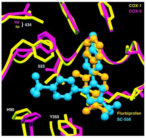
Isoform-selective inhibitor binding. The COX-1 and COX-2 active sites are shown superimposed (COX-1, yellow; COX-2, pink). Two inhibitors are seen: flurbiprofen (orange), a nonselective inhibitor, and SC-558 (blue), a COX-2–selective inhibitor. NSAIDs achieve COX inhibition by occupying the upper portion of the active site channel, preventing the fatty acid substrate from gaining access to the active site tyrosine seen at the upper right. Note how the COX-2–selective inhibitor projects leftward into a side pocket that is not exploited by the nonselective inhibitor.
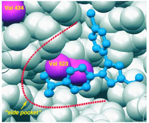
A view of the “side pocket” found at the side of the COX-2 active site. The COX-2–selective inhibitor SC-558 is shown in blue, bound in the active site. Protein residues are shown as van der Waals spheres; valine 434 and valine 523 are shown in pink. In COX-1, both these residues are isoleucines; the additional bulk contributed by the two extra methyl groups is sufficient to close down this small alcove so that no side pocket is found in COX-1.
References
Publication types
MeSH terms
Substances
LinkOut - more resources
Full Text Sources
Research Materials
