

Review
doi: 10.1172/JCI13210.
Arachidonic acid as a bioactive molecule
Affiliations
- PMID: 11390413
- PMCID: PMC209328
- DOI: 10.1172/JCI13210
Item in Clipboard
Review
Arachidonic acid as a bioactive molecule
J Clin Invest.
2001 Jun.
No abstract available
Figures
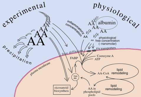
Arachidonic acid concentrations in experimental and physiological settings. Differing sizes of the AA (arachidonic acid) represent differing concentrations. The concentrations range from the nanomolar level for extracellular, unbound, free AA in the physiological setting (right side), up to the high micromolar levels, often used in the absence of binding proteins, in the experimental situation (left side). The ranges on concentrations in practice are considerably more divergent than are represented here by the size of AA. Proteins in the cell membrane may facilitate uptake of extracellular arachidonate and its presentation to a coenzyme A synthetase for esterification. Fatty acid binding proteins (FABP) facilitate transfer and modulate the available concentrations of arachidonic acid within cells. FA transporter = fatty acid transporter.
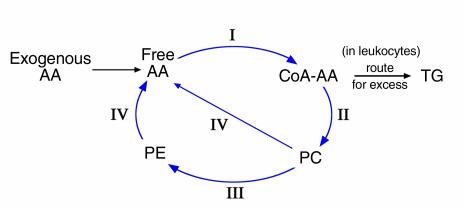
A simplified overview of arachidonate esterification and phospholipid remodeling in cells. The key enzymes are (I) Acyl Coenzyme A synthetase (or ligase), (II) PC/CoA-AA, phosphatidylcholine/CoA arachidonate transacylase, (III) CoA-IT, CoA-independent transacylase, and (IV) iPLA2, calcium-independent PLA2. Not shown are distinct pools of free arachidonic acid that may be generated in subcellular compartments by the actions of other lipases (such as secreted PLA2 [sPLA2], cytosolic PLA2 [cPLA2], phospholipases C or D [PLC, PLD]), e.g. on the nuclear membrane for leukotriene biosynthesis. AA, arachidonic acid; TG, triglyceride.
References
-
- Bild GS, Ramadoss CS, Axelrod B. Effect of solvent polarity on the activity of soybean lipoxygenase isozymes. Lipids. 1977;12:732–735.
-
- Glickman MH, Klinman JP. Nature of rate-limiting steps in the soybean lipoxygenase-1 reaction. Biochemistry. 1995;34:14077–14092. - PubMed
-
- Spector AA. Structure and lipid binding properties of serum albumin. J Lipid Res. 1986;128:320–329. - PubMed
-
- McArthur MJ, et al. Cellular uptake and intracellular trafficking of long chain fatty acids. J Lipid Res. 1999;40:1371–1383. - PubMed
-
- Berk PD, Stump DD. Mechanisms of cellular uptake of long chain free fatty acids. Mol Cell Biochem. 1999;192:17–31. - PubMed
Publication types
MeSH terms
Substances
Grants and funding
LinkOut - more resources
Full Text Sources
Other Literature Sources
