. 2001 Jun 4;193(11):1295-302.
doi: 10.1084/jem.193.11.1295.

Human cd25(+)cd4(+) t regulatory cells suppress naive and memory T cell proliferation and can be expanded in vitro without loss of function

Affiliations

Human cd25(+)cd4(+) t regulatory cells suppress naive and memory T cell proliferation and can be expanded in vitro without loss of function

M K Levings et al. J Exp Med. .

Abstract

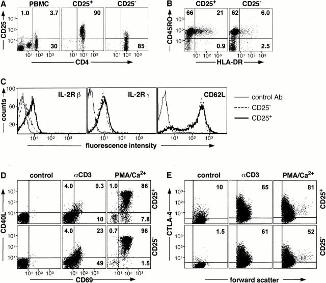
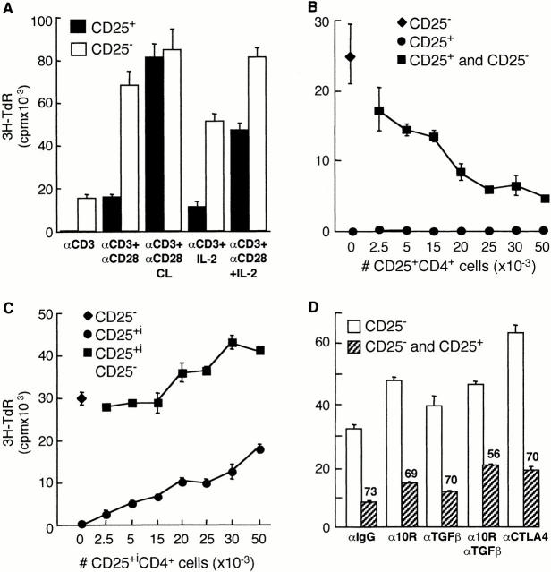
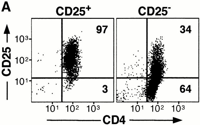
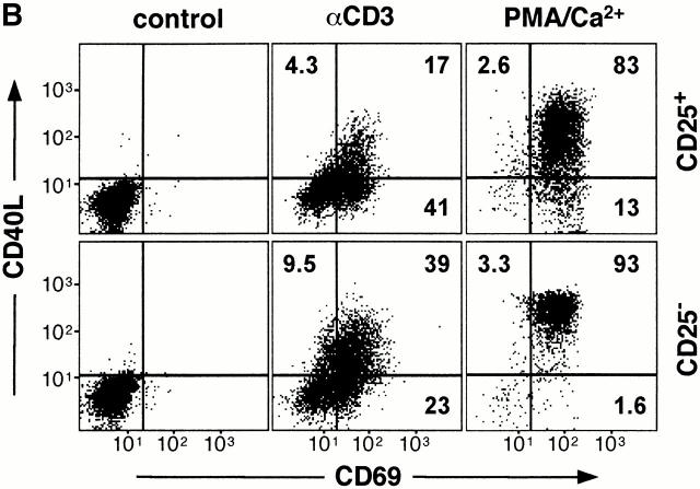
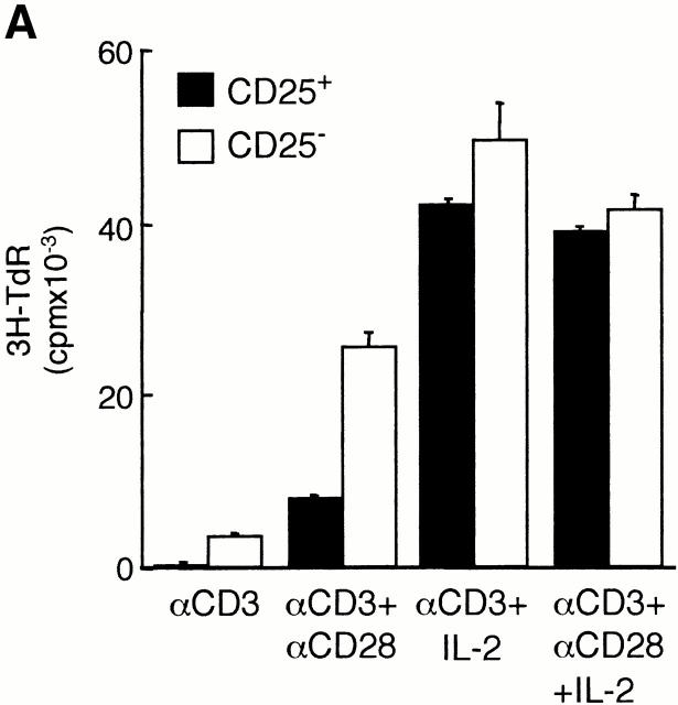
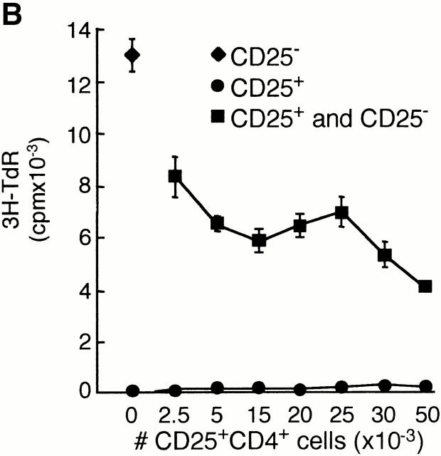
Active suppression by T regulatory (Tr) cells plays an important role in the downregulation of T cell responses to foreign and self-antigens. Mouse CD4(+) Tr cells that express CD25 possess remarkable suppressive activity in vitro and in autoimmune disease models in vivo. Thus far, the existence of a similar subset of CD25(+)CD4(+) Tr cells in humans has not been reported. Here we show that human CD25(+)CD4(+) Tr cells isolated from peripheral blood failed to proliferate and displayed reduced expression of CD40 ligand (CD40L), in response to T cell receptor-mediated polyclonal activation, but strongly upregulated cytotoxic T lymphocyte-associated antigen (CTLA)-4. Human CD25(+)CD4(+) Tr cells also did not proliferate in response to allogeneic antigen-presenting cells, but they produced interleukin (IL)-10, transforming growth factor (TGF)-beta, low levels of interferon (IFN)-gamma, and no IL-4 or IL-2. Importantly, CD25(+)CD4(+) Tr cells strongly inhibited the proliferative responses of both naive and memory CD4(+) T cells to alloantigens, but neither IL-10, TGF-beta, nor CTLA-4 seemed to be directly required for their suppressive effects. CD25(+)CD4(+) Tr cells could be expanded in vitro in the presence of IL-2 and allogeneic feeder cells and maintained their suppressive capacities. These findings that CD25(+)CD4(+) Tr cells with immunosuppressive effects can be isolated from peripheral blood and expanded in vitro without loss of function represent a major advance towards the therapeutic use of these cells in T cell-mediated diseases.


Figures

Figure 1
Figure 1
Isolation and cell surface phenotype of human CD25+CD4+ Tr cells. CD4+ T cells were isolated from PBMCs and separated into CD25+ and CD25 fractions. Purity (A) and expression of CD45RO, HLA-DR (B), IL-2Rβ, IL-2Rγ, and CD62L (C) was determined by FACS®. CD25+CD4+ and CD25CD4+ T cells were cultured either in medium alone or activated with immobilized anti-CD3 mAbs or PMA and calcium ionophore for 6 (D) or 24 h (E). Cells were analyzed for surface expression of CD40L and CD69 (D) and for intracytoplasmic expression of CTLA-4 (E). Results are representative of six independent experiments.
Figure 2
Figure 2
CD25+CD4+ Tr cells are anergic and suppress proliferation to alloantigens. Purified CD25+CD4+ Tr cells (100,000 cells per well) were tested for their ability to proliferate in response to immobilized anti-CD3 mAbs (αCD3; 10 μg/ml) in the absence or presence of soluble anti-CD28 mAbs (αCD28; 1 μg/ml), secondary rabbit anti–mouse Abs (αCD28 CL; 10 μg/ml), and/or 100 U/ml IL-2. After 72 h of culture, [3H]thymidine was added for an additional 16 h (A). CD25CD4+ T cells (50,000 cells per well) were tested for their ability to proliferate in response to allogeneic APCs in the absence or presence of increasing numbers of autologous CD25+CD4+ Tr cells (B). CD25CD4+ T cells were activated to induce CD25 expression. After 48 h, T cells that became CD25+ (CD25+i) were purified and tested for their ability to suppress proliferation of CD25 T cells in response to alloantigens (C). CD25CD4+ T cells were activated by alloantigens with or without CD25+CD4+ Tr cells (1:1 ratio) in the presence of the indicated mAbs (10 μg/ml). Numbers represent the percent reduction in proliferation compared with culture in the absence of CD25+CD4+ Tr cells (D). For B–D, after 96 h, [3H]thymidine was added for an additional 16 h. Results are representative of six independent experiments for A, nine for B, and three for C and D.
Figure 3
Figure 3
Expansion and cell surface phenotype of CD25+CD4+ Tr cells. CD25+ and CD25CD4+ T cells were purified and activated with anti-CD3 mAbs, allogeneic feeder cell mixture, and exogenous IL-2. Cells were split as necessary and after 2 wk were analyzed by FACS® for expression of CD25 and CD4 (A). In parallel, cells were analyzed for cell surface expression of CD40L and CD69 after activation for 6 h with immobilized anti-CD3 mAbs or PMA and calcium ionophore (B). Constitutive levels of CTLA-4 expression was determined by intracytoplasmic staining (C). Results are representative of three independent experiments.
Figure 3
Figure 3
Expansion and cell surface phenotype of CD25+CD4+ Tr cells. CD25+ and CD25CD4+ T cells were purified and activated with anti-CD3 mAbs, allogeneic feeder cell mixture, and exogenous IL-2. Cells were split as necessary and after 2 wk were analyzed by FACS® for expression of CD25 and CD4 (A). In parallel, cells were analyzed for cell surface expression of CD40L and CD69 after activation for 6 h with immobilized anti-CD3 mAbs or PMA and calcium ionophore (B). Constitutive levels of CTLA-4 expression was determined by intracytoplasmic staining (C). Results are representative of three independent experiments.
Figure 4
Figure 4
Cultured CD25+CD4+ Tr cells retain their suppressive capacity. CD25+ and CD25CD4+ T cells were purified and activated with anti-CD3 mAbs, allogeneic feeder cell mixture, and exogenous IL-2. After 14 d of culture, T cells were tested for their ability to proliferate in response to 10 μg/ml anti-CD3 mAbs in the absence or presence of 1 μg/ml soluble anti-CD28 mAbs and/or 100 U/ml IL-2 (A). Cultured CD25CD4+ T cells (50,000 cells per well) were tested for their ability to proliferate in response to alloantigens in the absence or presence of increasing numbers of in vitro–cultured, autologous CD25+CD4+ Tr cells (B). Cultured CD25CD4+ T cells were activated by allogeneic APCs (from a donor different from that used for expansion) with or without CD25+CD4+ Tr cells (1:1 ratio) in the presence of the indicated mAbs (10 μg/ml). Numbers represent the percent reduction in proliferation compared with culture in the absence of CD25+CD4+ Tr cells (C). For all cultures, after 48 h, [3H]thymidine was added for an additional 16 h. Results are representative of three independent experiments.
Figure 4
Figure 4
Cultured CD25+CD4+ Tr cells retain their suppressive capacity. CD25+ and CD25CD4+ T cells were purified and activated with anti-CD3 mAbs, allogeneic feeder cell mixture, and exogenous IL-2. After 14 d of culture, T cells were tested for their ability to proliferate in response to 10 μg/ml anti-CD3 mAbs in the absence or presence of 1 μg/ml soluble anti-CD28 mAbs and/or 100 U/ml IL-2 (A). Cultured CD25CD4+ T cells (50,000 cells per well) were tested for their ability to proliferate in response to alloantigens in the absence or presence of increasing numbers of in vitro–cultured, autologous CD25+CD4+ Tr cells (B). Cultured CD25CD4+ T cells were activated by allogeneic APCs (from a donor different from that used for expansion) with or without CD25+CD4+ Tr cells (1:1 ratio) in the presence of the indicated mAbs (10 μg/ml). Numbers represent the percent reduction in proliferation compared with culture in the absence of CD25+CD4+ Tr cells (C). For all cultures, after 48 h, [3H]thymidine was added for an additional 16 h. Results are representative of three independent experiments.

References

    1. Roncarolo M.G., Levings M.K. The role of different subsets of T regulatory cells in controlling autoimmunity. Curr. Opin. Immunol. 2000;12:676–683. - PubMed
    1. Sakaguchi S. Regulatory T cellskey controllers of immunologic self-tolerance. Cell. 2000;101:455–458. - PubMed
    1. Shevach E.M. Regulatory T cells in autoimmmunity. Annu. Rev. Immunol. 2000;18:423–449. - PubMed
    1. Cottrez F., Hurst S.D., Coffman R.L., Groux H. T regulatory cells 1 inhibit a Th2-specific response in vivo. J. Immunol. 2000;165:4848–4853. - PubMed
    1. Asseman C., Mauze S., Leach M.W., Coffman R.L., Powrie F. An essential role for interleukin 10 in the function of regulatory T cells that inhibit intestinal inflammation. J. Exp. Med. 1999;190:995–1004. - PMC - PubMed

Publication types