. 2001 Jul;75(13):6183-92.
doi: 10.1128/JVI.75.13.6183-6192.2001.

Varicella-zoster virus infection of human dendritic cells and transmission to T cells: implications for virus dissemination in the host

Affiliations

Varicella-zoster virus infection of human dendritic cells and transmission to T cells: implications for virus dissemination in the host

A Abendroth et al. J Virol. 2001 Jul.

Abstract

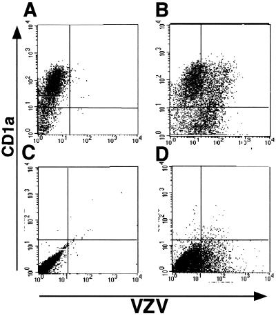
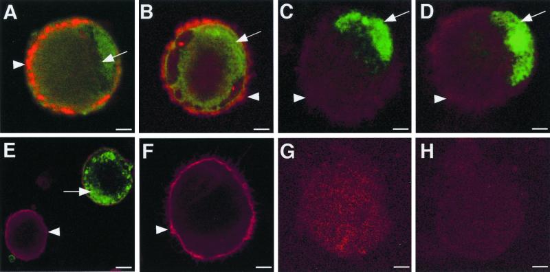
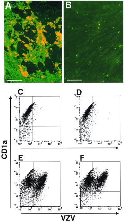
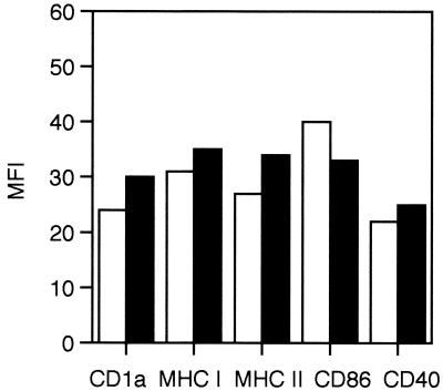
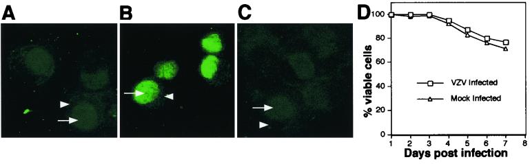
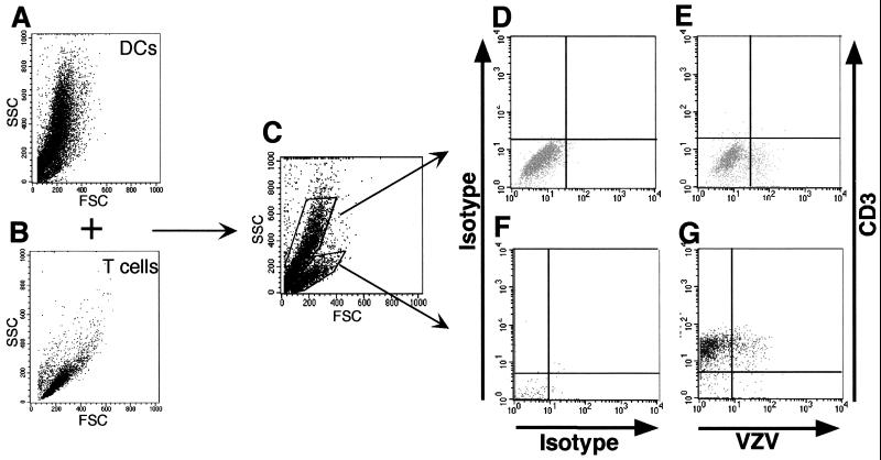
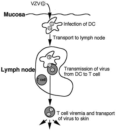
During primary varicella-zoster virus (VZV) infection, it is presumed that virus is transmitted from mucosal sites to regional lymph nodes, where T cells become infected. The cell type responsible for VZV transport from the mucosa to the lymph nodes has not been defined. In this study, we assessed the susceptibility of human monocyte-derived dendritic cells to infection with VZV. Dendritic cells were inoculated with the VZV strain Schenke and assessed by flow cytometry for VZV and dendritic cell (CD1a) antigen expression. In five replicate experiments, 34.4% +/- 6.6% (mean +/- SEM) of CD1a(+) cells were also VZV antigen positive. Dendritic cells were also shown to be susceptible to VZV infection by the detection of immediate-early (IE62), early (ORF29), and late (gC) gene products in CD1a(+) dendritic cells. Infectious virus was recovered from infected dendritic cells, and cell-to-cell contact was required for transmission of virus to permissive fibroblasts. VZV-infected dendritic cells showed no significant decrease in cell viability or evidence of apoptosis and did not exhibit altered cell surface levels of major histocompatibility complex (MHC) class I, MHC class II, CD86, CD40, or CD1a. Significantly, when autologous T lymphocytes were incubated with VZV-infected dendritic cells, VZV antigens were readily detected in CD3(+) T lymphocytes and infectious virus was recovered from these cells. These data provide the first evidence that dendritic cells are permissive to VZV and that dendritic cell infection can lead to transmission of virus to T lymphocytes. These findings have implications for our understanding of how virus may be disseminated during primary VZV infection.


Figures

FIG. 1
FIG. 1
Flow cytometry analysis of expression of VZV antigen on the surfaces of human DCs and human fibroblasts infected with VZV. Human DCs were inoculated with uninfected or VZV-infected fibroblasts and collected 2 days postinoculation. Cells from mock-infected DCs (A), VZV-infected DCs (B), uninfected fibroblasts (C), and VZV-infected fibroblasts (D) were stained with antibodies and fluorescent conjugates to CD1a and VZV proteins and analyzed by flow cytometry.
FIG. 2
FIG. 2
Immunofluorescence staining of CD1a and VZV antigens in VZV-infected DCs. Two days postinfection, DCs infected with VZV strain Schenke (A to E, G) and mock infected (F and H) were incubated with a mouse monoclonal antibody to CD1a (A to F) and rabbit polyclonal antibodies to ORF62 (A and F), ORF4 (B), ORF29 (C), ORF61 (D), and glycoprotein C (E). CD1a binding was detected using a Texas Red-conjugated anti-mouse antibody (red fluorescence). Rabbit antibodies generated against viral proteins were detected using a FITC-conjugated anti-rabbit antibody (green fluorescence). Negative controls were VZV-infected and mock-infected DCs incubated with isotype control antibodies (G and H, respectively). All negative control images were obtained by increasing the laser voltage to enable the visualization of cells. The arrowheads indicate CD1a staining, and the arrows indicate VZV-specific staining.
FIG. 3
FIG. 3
Infectious center assay and flow cytometry analysis of VZV-infected DCs. Dendritic cells inoculated with VZV-infected HFF were harvested at day 4 postinoculation. CD1a+ sorted cells (A) or media from VZV-infected fibroblasts (B) were incubated directly on HFF monolayers. Seven days later, cell monolayers were fixed and incubated with a human polyclonal VZV immune serum, followed by an FITC-conjugated anti-human antibody. In parallel, flow cytometry analysis of VZV antigen expression was performed on DCs stripped of surface antigens. Dendritic cells were inoculated with VZV-infected (E and F) or mock-infected (C and D) fibroblasts. Four days postinoculation, VZV-infected and mock-infected DCs from unstripped (C and E) and low-pH-buffer (stripped) cultures (D and F) were immunostained for CD1a and VZV antigens and analyzed by flow cytometry.
FIG. 4
FIG. 4
Mean fluorescence intensity of CD1a, MHC class I, MHC class II, CD86, and CD40 protein expression on VZV-infected DCs. VZV-infected DCs were harvested at day 2 postinfection and dual-stained with VZV immune polyclonal human serum and monoclonal antibodies specific for CD1a, MHC class I (MHC I), MHC class II (MHC II), CD86, or CD40. The mean fluorescence intensity (MFI) values of VZV cells (white boxes) and VZV+ cell populations (black boxes) are shown.
FIG. 5
FIG. 5
Assessment of apoptosis and cell viability in VZV-infected DCs. DCs inoculated with uninfected fibroblasts or infected fibroblasts were harvested and stained with trypan blue or spotted onto sides for a TUNEL assay. TUNEL staining on VZV-infected DCs (A), in the presence of DNase (B), and in the absence of TdT (C) is shown. Each arrowhead indicates the cytoplasm, and each arrow indicates the nucleus of the cell. The percentages of viable cells for infected and uninfected DC culture over a 7-day culture period are shown (D).
FIG. 6
FIG. 6
Flow cytometry analysis of VZV-infected DCs cultured with T lymphocytes. VZV-infected DCs and autologous T cells were analyzed for their FSC and SSC properties before (A and B) and after (C) coculturing. The cocultured cells were stained 2 days later with antibodies and fluorescent conjugates to CD3 and VZV proteins. The flow cytometry analyses for T lymphocytes (G) and DCs (E) are shown with their appropriate isotype controls (F and D). The same numbers of cells were stained with antigen-specific (VZV and CD3) and isotype control antibodies.
FIG. 7
FIG. 7
Immunofluorescence staining and flow cytometry analysis of human T lymphocytes inoculated with VZV-infected DCs. T lymphocytes were inoculated with uninfected or VZV-infected DCs. Two days later, cell preparations from mock-infected T cells (A, C, and E) and VZV-infected T cells (B, D, and F) were stained with antibodies and fluorescent conjugates to CD3, CD4, CD8, and VZV proteins and analyzed by confocal microscopy (A and B) or flow cytometry (C to F). The arrowhead indicates VZV-antigen-specific staining, and the arrows indicate CD3-specific staining.
FIG. 8
FIG. 8
Proposed model of virus transport from the site of mucosal inoculation to the T cells in lymph nodes during primary VZV infection.

References

    1. Abendroth A, Arvin A M. Varicella zoster virus immune evasion. Immunol Rev. 1999;168:143–156. - PubMed
    1. Abendroth A, Slobedman B, Eunice L, Mellins E, Wallace M, Arvin A M. Modulation of MHC class II protein expression by varicella-zoster virus. J Virol. 2000;74:1900–1907. - PMC - PubMed
    1. Arvin A M. Varicella zoster virus. In: Fields B N, Knipe D M, Howley P M, editors. Fields virology. New York, N.Y: Raven; 1996. pp. 2547–2585.
    1. Arvin A M, Moffat J F, Redman R. Varicella-zoster virus: aspects of pathogenesis and host response to natural infection and varicella vaccine. Adv Virus Res. 1996;46:263–309. - PubMed
    1. Arvin A M. Varicella zoster virus: virologic and immunologic aspects of persistent infection. In: Ahmed R, Chen I, editors. Persistent viral infections. New York, N.Y: John Wiley and Sons Ltd.; 1998. pp. 183–208.

Publication types