. 2001 May-Jun;8(3):148-55.
doi: 10.1101/lm.37601.

Contextual and auditory fear conditioning are mediated by the lateral, basal, and central amygdaloid nuclei in rats

Affiliations

Contextual and auditory fear conditioning are mediated by the lateral, basal, and central amygdaloid nuclei in rats

K A Goosens et al. Learn Mem. 2001 May-Jun.

Abstract

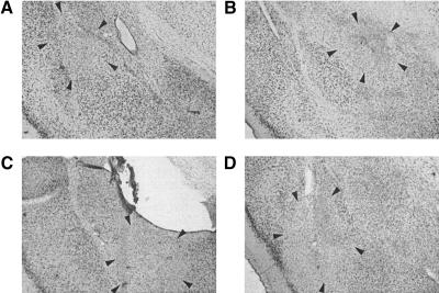
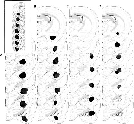
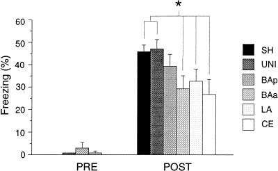
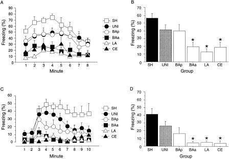
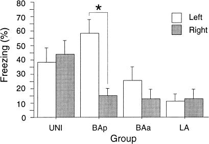
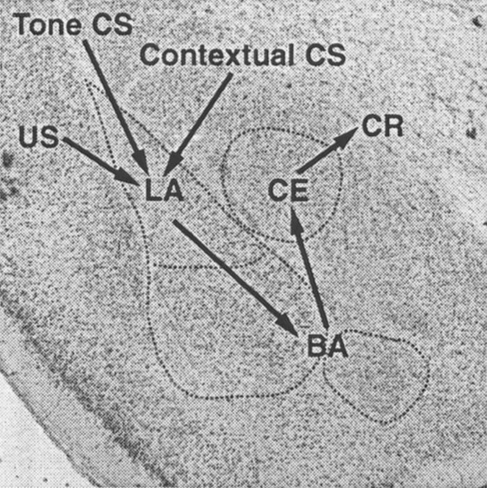
A large body of literature implicates the amygdala in Pavlovian fear conditioning. In this study, we examined the contribution of individual amygdaloid nuclei to contextual and auditory fear conditioning in rats. Prior to fear conditioning, rats received a large electrolytic lesion of the amygdala in one hemisphere, and a nucleus-specific neurotoxic lesion in the contralateral hemisphere. Neurotoxic lesions targeted either the lateral nucleus (LA), basolateral and basomedial nuclei (basal nuclei), or central nucleus (CE) of the amygdala. LA and CE lesions attenuated freezing to both contextual and auditory conditional stimuli (CSs). Lesions of the basal nuclei produced deficits in contextual and auditory fear conditioning only when the damage extended into the anterior divisions of the basal nuclei; damage limited to the posterior divisions of the basal nuclei did not significantly impair conditioning to either auditory or contextual CS. These effects were typically not lateralized, although neurotoxic lesions of the posterior divisions of the basal nuclei had greater effects on contextual fear conditioning when the contralateral electrolytic lesion was placed in the right hemisphere. These results indicate that there is significant overlap within the amygdala in the neural pathways mediating fear conditioning to contextual and acoustic CS, and that these forms of learning are not anatomically dissociable at the level of amygdaloid nuclei.


Figures

Figure 1
Figure 1
Neurotoxic amygdala lesions. The photomicrographs are of thionin-stained coronal sections from the brains of rats with representative lesions of the (A) lateral nucleus of the amygdala (LA), (B) central nucleus of the amygdala (CE), and (C,D) basal amygdaloid nuclei (BAp in C, BAa in D).
Figure 2
Figure 2
Schematic representation of the minimum (gray) and maximum (black) extent of damage of unilateral excitotoxic lesions to the (A) posterior divisions of the basolateral and basomedial nuclei of the amygdala (BAp), (B) anterior divisions of the basolateral and basomedial nuclei of the amygdala (BAa), (C) lateral nucleus of the amygdala (LA), and (D) central nucleus of the amygdala (CE). The extent of unilateral electrolytic lesions of the amygdala is depicted in the inset. Brain images are adapted from Swanson (1999).
Figure 3
Figure 3
Conditional freezing during training. Mean (±SEM) percentage of freezing in the 3 min prior to receiving tone–footshock pairings (PRE) and mean (±SEM) percentage of freezing for the postshock period in which freezing was maximal (POST). Asterisk indicates that P < 0.05, in planned comparisons between the UNI and SHAM groups and groups with discrete neurotoxic lesions of amygdaloid nuclei.
Figure 4
Figure 4
Pretraining neurotoxic lesions of amygdaloid nuclei and conditional freezing. (A) Mean (±SEM) percentage of freezing during an 8-min extinction in the conditioning context. (B) Mean (±SEM) percentage of freezing averaged across the 8-min context extinction test. (C) Mean (±SEM) percentage of freezing during an 8-min tone extinction test in the B context. The tone was present during minutes 3 through 10. (D) Mean (±SEM) percentage of freezing averaged across the 8-min tone test. Asterisk indicates that P < 0.05, in planned comparisons between the UNI group and groups with discrete neurotoxic lesions of amygdaloid nuclei.
Figure 5
Figure 5
Hemisphere of the electrolytic amygdala lesion and contextual conditional freezing. These data are adapted from Figure 3B, with each group divided according to the hemisphere in which the large electrolytic lesion of the total amygdala was placed. Each group has a minimum of five rats. The CE group was not included in this analysis because there were too few rats to meet the group minimum. Asterisk indicates that P < 0.05, in planned comparisons.
Figure 6
Figure 6
Anatomical model of fear conditioning circuits within the amygdala. Contextual and auditory CS converge on LA neurons, where they come into association with the footshock US. Indirect projections from the LA to the CE via the basal nuclei mediate expression of both tone–US and context–US associations.

References

    1. Amorapanth P, LeDoux JE, Nader K. Different lateral amygdala outputs mediate reactions and actions elicited by a fear-arousing stimulus. Nature Neurosci. 2000;3:74–79. - PubMed
    1. Cahill L, Haier RJ, Fallon J, Alkire MT, Tang C, Keator D, Wu J, McGaugh JL. Amygdala activity at encoding correlated with long-term free recall of emotional information. Proc Natl Acad Sci. 1996;93:8016–8021. - PMC - PubMed
    1. Campeau S, Davis M. Involvement of the central nucleus and basolateral complex of the amygdala in fear conditioning measured with fear-potentiated startle in rats trained concurrently with auditory and visual conditioned stimuli. J Neurosci. 1995;15:2301–2311. - PMC - PubMed
    1. Canteras NS, Swanson LW. Projections of the ventral subiculum to the amygdala, septum, and hypothalamus: A PHAL anterograde tract-tracing study in the rat. J Comp Neurol. 1992;324:180–194. - PubMed
    1. Coleman-Mesches K, McGaugh JL. Differential effects of pretraining inactivation of the right or left amygdala on retention of inhibitory avoidance training. Behav Neurosci. 1995;109:642–647. - PubMed

Publication types

LinkOut - more resources