. 2001 Jul;21(13):4369-78.
doi: 10.1128/MCB.21.13.4369-4378.2001.

Complex functions of AP-1 transcription factors in differentiation and survival of PC12 cells

Affiliations

Complex functions of AP-1 transcription factors in differentiation and survival of PC12 cells

S Leppä et al. Mol Cell Biol. 2001 Jul.

Abstract

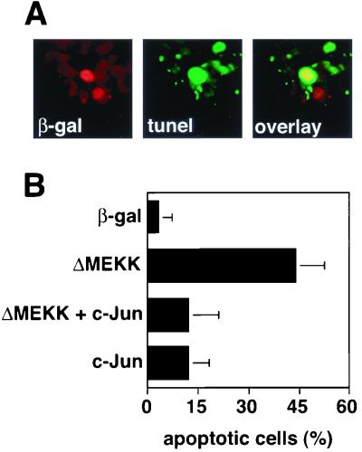
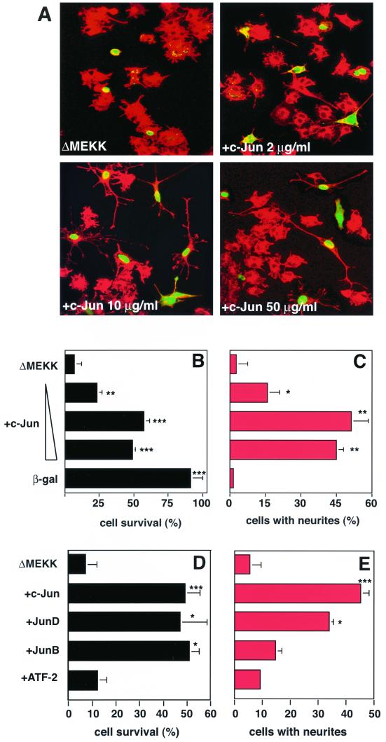
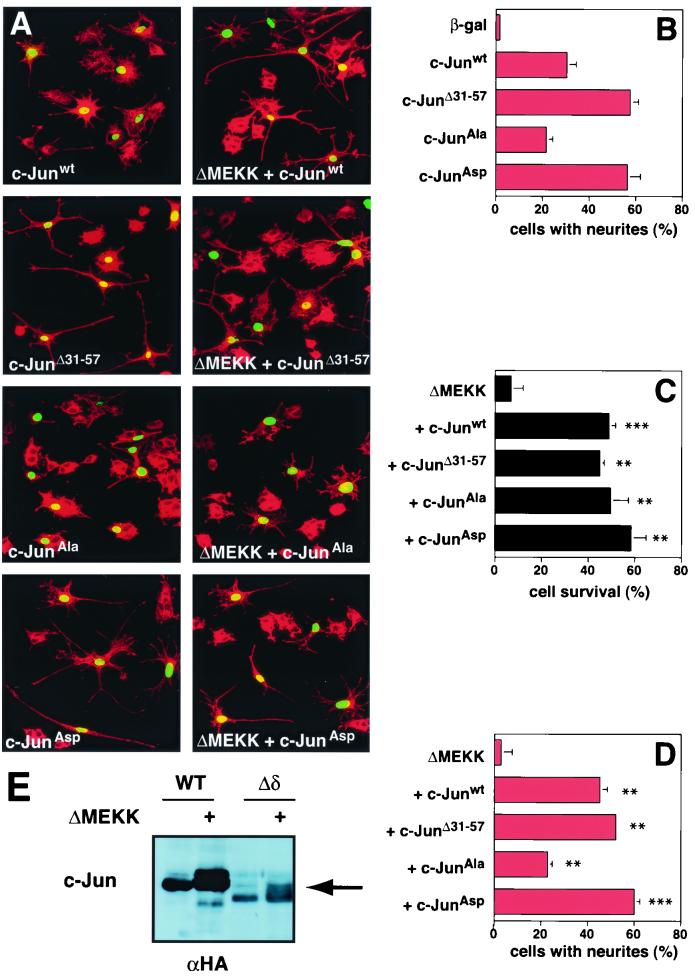
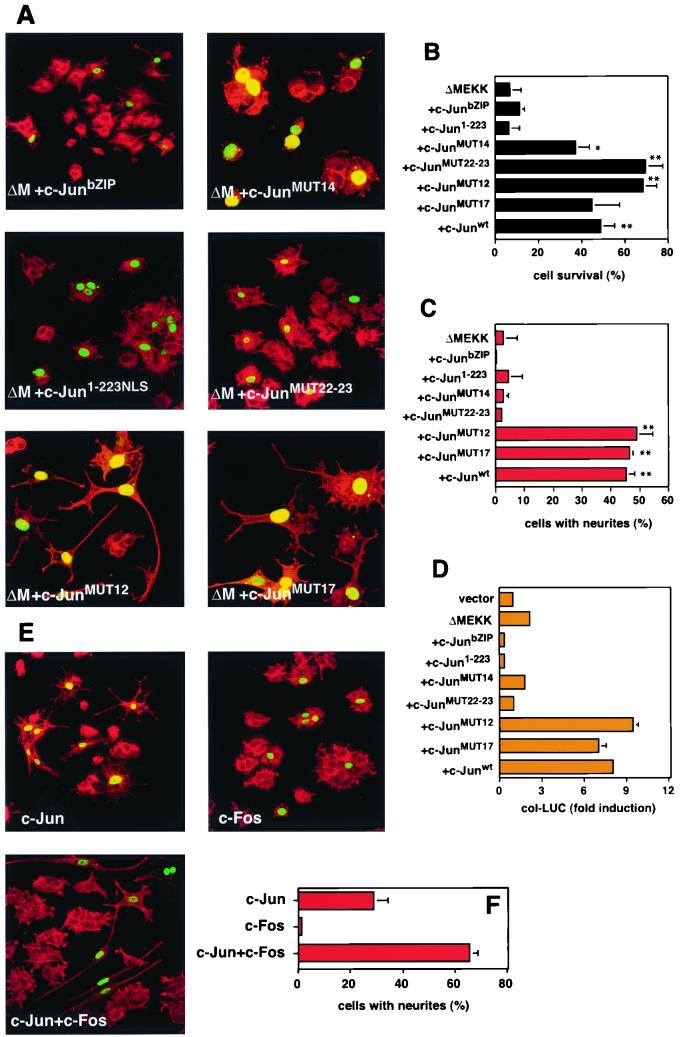
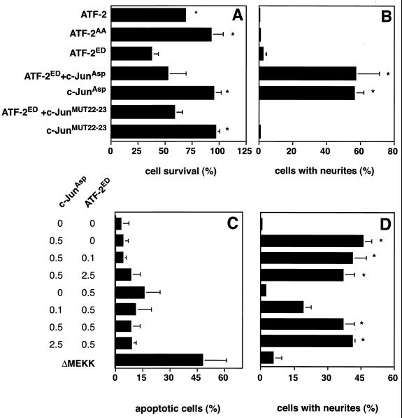
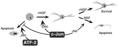
c-Jun activation by mitogen-activated protein kinases has been implicated in various cellular signal responses. We investigated how JNK and c-Jun contribute to neuronal differentiation, cell survival, and apoptosis. In differentiated PC12 cells, JNK signaling can induce apoptosis and c-Jun mediates this response. In contrast, we show that in PC12 cells that are not yet differentiated, the AP-1 family member ATF-2 and not c-Jun acts as an executor of apoptosis. In this context c-Jun expression protects against apoptosis and triggers neurite formation. Thus, c-Jun has opposite functions before and after neuronal differentiation. These findings suggest a model in which the balance between ATF-2 and Jun activity in PC12 cells governs the choice between differentiation towards a neuronal fate and an apoptotic program. Further analysis of c-Jun mutants showed that the differentiation response requires functional dimerization and DNA-binding domains and that it is stimulated by phosphorylation in the transactivation domain. In contrast, c-Jun mutants incompetent for DNA binding or dimerization and also mutants lacking JNK binding and phosphorylation sites that cannot elicit neuronal differentiation efficiently protect PC12 cells from apoptosis. Hence, the protective role of c-Jun appears to be mediated by an unconventional mechanism that is separable from its function as a classical AP-1 transcription factor.


Figures

FIG. 1
FIG. 1
c-Jun expression rescues PC12 cells from ΔMEKK-induced apoptosis. (A) Induction of apoptosis in PC12 cells expressing ΔMEKK. PC12 cells were transfected with expression vectors for ΔMEKK and nuclear β-galactosidase (β-gal). After 24 h, the cells were fixed and double stained with anti-β-galactosidase antibody to detect transfected cells and with TUNEL reagent to mark cells undergoing apoptosis. Nuclei of cells expressing ΔMEKK appear red (left panel), and apoptotic cells are visualized in green (middle panel). (B) Quantification of apoptosis. The percentage of TUNEL-positive cells among the transfected cells was determined. The data are the means ± standard errors of two separate experiments.
FIG. 2
FIG. 2
Jun proteins but not ATF-2 rescue PC12 cells from ΔMEKK-induced apoptosis. (A) Morphology of PC12 cells expressing ΔMEKK alone or ΔMEKK with increasing concentrations of c-Jun, as indicated. Nuclear β-galactosidase was coexpressed to mark the injected cells. After 36 h, the cells were fixed and stained with anti-β-galactosidase. Injected cells were detected using FITC-labeled secondary antibodies (green), and the morphology of the cells was visualized by actin staining (red). Cells were examined by confocal microscopy. (B and D) Quantification of cell survival. The percentage of viable cells was determined. In apoptotic cells, β-galactosidase staining is punctate, and the cell bodies have shrunk. (C and E) Quantitation of neurite outgrowth. The percentage of the cells with neurites exceeding twice the cell length among the microinjected (FITC positive) cells is shown. c-Jun, JunB, JunD, and ATF-2 were compared for their ability to counteract ΔMEKK-induced cell death (D) and to induce neuronal differentiation (E). The data are the means ± standard errors of two or three separate experiments. Statistically significant differences from values for ΔMEKK-expressing cells are indicated as follows: ∗, P < 0.05; ∗∗, P < 0.01; ∗∗∗, P < 0.001.
FIG. 3
FIG. 3
c-Jun-mediated protection from ΔMEKK-induced apoptosis is JNK independent. (A) Morphology of cells expressing c-Jun derivatives and ΔMEKK. PC12 cells were injected with c-Junwt, c-JunΔ31–57, c-JunAla, or c-JunAsp expression vectors in the presence or absence of ΔMEKK, as indicated. Nuclear β-galactosidase was coexpressed to mark the injected cells. The cells were fixed after 40 h, stained with anti-β-galactosidase (green) and TRITC-phalloidin (red), and examined by confocal microscopy. (B to D) Quantification of cell survival and neurite outgrowth was performed as for Fig. 1. The data are means ± standard errors of two or three separate experiments. Statistically significant differences from values for ΔMEKK-expressing cells are indicated as follows: ∗, P < 0.05; ∗∗, P < 0.01; ∗∗∗, P < 0.001. (E) c-JunΔ31–57 is phosphorylated in response to ΔMEKK. HA-tagged c-Jun and c-JunΔ31–57 were expressed alone or with ΔMEKK in 293 cells. Cells were harvested 24 h posttransfection, and whole-cell extracts were analyzed by SDS-polyacrylamide gel electrophoresis and immunoblotting using anti-HA antibody.
FIG. 4
FIG. 4
c-Jun dimerization and DNA binding are not necessary for rescue from ΔMEKK-induced apoptosis. (A) Morphology of cells expressing c-Jun mutants and ΔMEKK. PC12 cells were injected with expression vectors for ΔMEKK together with c-Junwt or various c-Jun mutants as indicated. A plasmid coding for nuclear β-galactosidase was coexpressed to mark the injected cells. After 40 h, the cells were fixed, stained with anti-β-galactosidase and TRITC-phalloidin, and examined by confocal microscopy. MUT14 (K268I C269D) and MUT22-23 (L294P L308A) are defective in DNA binding and dimerization, respectively. MUT12 (K254I A255D) binds DNA poorly as a homodimer but well as a heterodimer with Fos. MUT17 (K288I A289D) forms slightly more stable homodimers than wild-type c-Jun. All mutants have been described previously (2). (B and C) Quantification of cell survival and neurite outgrowth was performed as for Fig. 1. The data are means ± standard errors of two or three separate experiments. Statistically significant differences from ΔMEKK-expressing cells are indicated as follows: ∗, P < 0.05; ∗∗, P < 0.01; ∗∗∗, P < 0.001. (D) Transcriptional activation of collagenase reporter by ΔMEKK and c-Jun mutants. PC12 cells were transfected with AP-1-responsive collagenase luciferase (col-LUC) and a control reporter (Renilla luciferase vector driven by the human ubiquitin promoter) together with expression vectors for ΔMEKK and c-Jun mutants, as indicated. The cells were harvested after 36 h for a dual luciferase assay. The firefly luciferase activity was normalized against the Renilla luciferase readings from the cotransfected internal control reporter. The data are representative of three independent experiments done in duplicate (means ± standard errors). (E) Morphology of cells expressing c-Jun and c-Fos. PC12 cells were injected with expression vectors for c-Jun, c-Fos, or both, as indicated. A plasmid coding for nuclear β-galactosidase was coexpressed to mark the injected cells. After 40 h, the cells were fixed and stained with anti-β-galactosidase and TRITC-phalloidin and examined by confocal microscopy. (F) Quantification of neurite outgrowth was performed as for Fig. 1. The data are the means ± standard errors of two or three separate experiments.
FIG. 5
FIG. 5
Activated ATF-2 induces cell death in PC12 cells. (A and B) PC12 cells were injected with plasmids encoding ATF-2WT, ATF-2AA, or ATF-2ED alone or in the presence of c-JunAsp or MUT22-23, as indicated. Nuclear β-galactosidase was coexpressed to mark the injected cells. (C and D) PC12 cells were transfected with 0.5 μg of the expression vectors for c-JunAsp or ATF-2ED per 3-cm dish in the presence of increasing amounts of ATF-2ED or c-JunAsp vectors (0, 0.1, 0.5, and 2.5 μg), respectively. Nuclear β-galactosidase was coexpressed. The cells were fixed after 48 h, stained with anti-β-galactosidase and TRITC-phalloidin, and examined by confocal microscopy. Quantification of cell survival and neurite outgrowth was performed as for Fig. 1. For panel C, cells undergoing apoptosis were identified and quantitated by measuring nuclear fragmentation with Hoechst staining. The data are the mean vs ± standard errors of two separate experiments. Statistically significant differences relative to values for ATF-2ED-expressing cells are indicated with an asterisk (P < 0.05).
FIG. 6
FIG. 6
Multiple functions of c-Jun in the control of apoptosis and neuronal differentiation. c-Jun prevents undifferentiated PC12 cells from undergoing MEKK1- and ATF-2-mediated apoptosis and promotes their neuronal differentiation. The first function does not require dimerization, DNA binding, or MAPK phosphorylation, whereas the latter appears to be a conventional AP-1 effect stimulated by ERK. Once differentiated, PC12 cells react to AP-1 activation through the JNK pathway by initiating apoptosis.

References

    1. Behrens A, Sibilia M, Wagner E F. Amino-terminal phosphorylation of c-Jun regulates stress-induced apoptosis and cellular proliferation. Nat Genet. 1999;21:326–329. - PubMed
    1. Bohmann D, Tjian R. Biochemical analysis of transcriptional activation by Jun: differential activity of c- and v-Jun. Cell. 1989;59:709–717. - PubMed
    1. Bos T J, Monteclaro F S, Mitsunobu F, Ball A R, Chang C H W, Nishimura T, Vogt P K. Efficient transformation of chicken embryo fibroblasts by c-Jun requires structural modifications in coding and noncoding sequences. Genes Dev. 1990;4:1677–1687. - PubMed
    1. Davis R J. Signal transduction by the JNK group of MAP kinases. Cell. 2000;103:239–252. - PubMed
    1. Dérijard B, Hibi M, Wu I, Barrett T, Su B, Deng T, Karin M, Davis R. JNK1: a protein kinase stimulated by UV-light and Ha-Ras that binds and phosphorylates the c-Jun activation domain. Cell. 1994;76:1025–1037. - PubMed

Publication types

MeSH terms

Substances