. 2001 Jul;21(13):4413-24.
doi: 10.1128/MCB.21.13.4413-4424.2001.

Human pex19p binds peroxisomal integral membrane proteins at regions distinct from their sorting sequences

Affiliations

Human pex19p binds peroxisomal integral membrane proteins at regions distinct from their sorting sequences

M Fransen et al. Mol Cell Biol. 2001 Jul.

Abstract

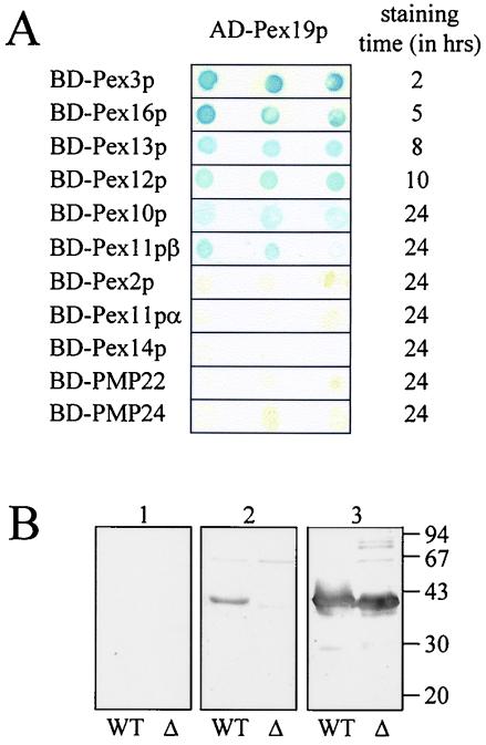
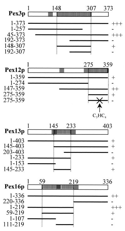
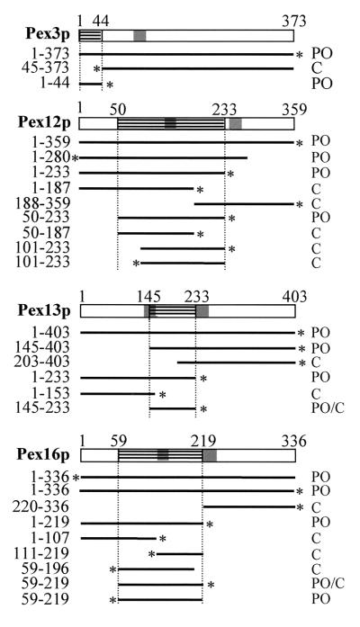
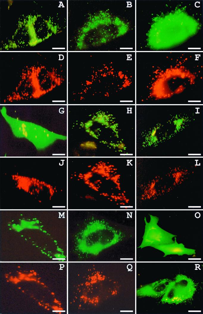
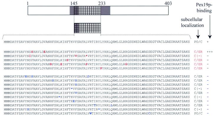
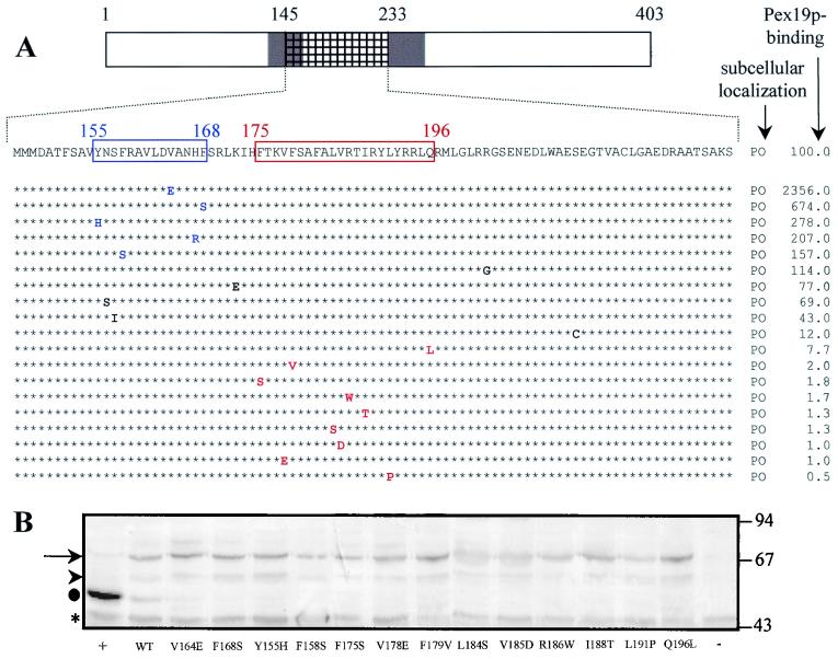
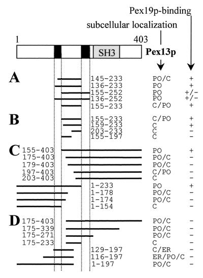
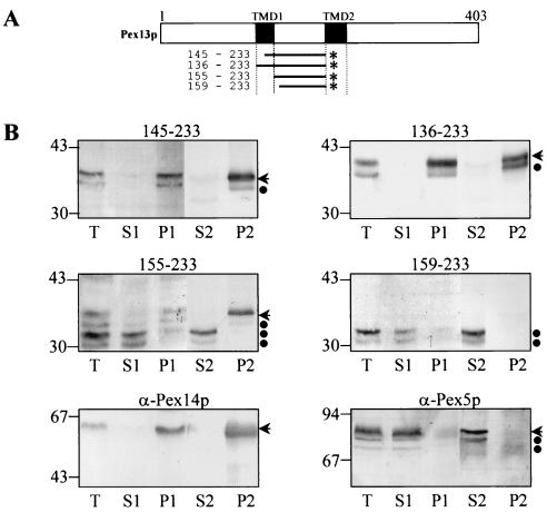
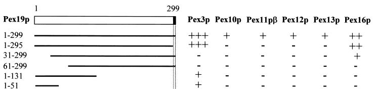
The molecular machinery underlying peroxisomal membrane biogenesis is not well understood. The observation that cells deficient in the peroxins Pex3p, Pex16p, and Pex19p lack peroxisomal membrane structures suggests that these molecules are involved in the initial stages of peroxisomal membrane formation. Pex19p, a predominantly cytosolic protein that can be farnesylated, binds multiple peroxisomal integral membrane proteins, and it has been suggested that it functions as a soluble receptor for the targeting of peroxisomal membrane proteins (PMPs) to the peroxisome. An alternative view proposes that Pex19p functions as a chaperone at the peroxisomal membrane. Here, we show that the peroxisomal sorting determinants and the Pex19p-binding domains of a number of PMPs are distinct entities. In addition, we extend the list of peroxins with which human Pex19p interacts to include the PMP Pex16p and show that Pex19p's CaaX prenylation motif is an important determinant in the affinity of Pex19p for Pex10p, Pex11pbeta, Pex12p, and Pex13p.


Figures

FIG. 1
FIG. 1
Pex19p interacts with multiple integral PMPs. (A) Human Pex19p, fused to the Gal4p AD, was tested for interaction with all known mammalian integral membrane peroxins, as well as with PMP22 and PMP24 fused to the Gal4p DNA-BD in the yeast two-hybrid system. Double transformants were selected and tested for β-galactosidase expression by using a filter assay with X-Gal as the substrate. Three representative independent transformants are shown. (B) Two micrograms of purified His6-tagged Pex19p(1-299) (WT) and Pex19p(31-299) (Δ) was subjected to sodium dodecyl sulfate-polyacrylamide gel electrophoresis, transferred to nitrocellulose, and incubated with a bacterial lysate containing no recombinant protein (panel 1), with His6-Pex3p(44-373) (panel 2), or with an anti-Pex19p antiserum (panel 3). Pex19p-Pex3p complexes were visualized by using an anti-Pex3p antiserum (1, 2). The migration of the molecular mass markers (in kilodaltons) is indicated.
FIG. 2
FIG. 2
Mapping of the Pex19p-binding sites of Pex3p, Pex12p, Pex13p, and Pex16p. Deletion constructs of Pex3p, Pex12p, Pex13p, and Pex16p fused to Gal4p-BD were tested for interaction with Pex19p fused to the Gal4p-AD in the yeast two-hybrid system. Double transformants were selected and assayed for β-galactosidase activity by using a filter assay with X-Gal as the substrate. The colony staining times were less than 2 h (+++), less than 5 h (++), or less than 10 h (+) or the colonies did not stain at all (−). TMDs are shaded. The smallest delineated Pex19p-BDs are hatched with vertical lines. Residue numbers are on top and on the left. X, alterations of the Pex12p C3HC4 RING residues from C-1 to W at position 304 and C-2 to Q at position 307.
FIG. 3
FIG. 3
Mapping of the mPTS of Pex3p, Pex12p, Pex13p, and Pex16p. CHO cells were transiently transfected with plasmids expressing deletion fragments of Pex3p, Pex12p, Pex13p, and Pex16p N terminally or C terminally fused to GFP (*). After 24 h, the cells were processed for direct fluorescence and the subcellular localization of the GFP-fusion proteins was determined: peroxisome (PO), cytosol (C), and peroxisome-cytosol (PO/C). TMDs are shaded. Residue numbers are on top and on the left. The smallest delineated domains sufficient to target the GFP reporter protein to the peroxisomes are hatched with horizontal lines.
FIG. 4
FIG. 4
Targeting of Pex13p-GFP fusion proteins in CHO cells. CHO cells transiently transfected with plasmids expressing the peroxisomal marker protein DsRed-KSKL (D, E, F, J, K, L, P, Q) and Pex13p(1-403) (A), Pex13p(145-233) (B), Pex13p(155-233) (C), Pex13p(159-233) (G), Pex13p(1-403)V164E (H), Pex13p(1-403)L191P (I), Pex13p(136-233) (M), Pex13p(145-233)R186W, S214C (N), Pex13p(145-233)F158S, V164E (O), or Pex13p(116-197) (R) N terminally fused to GFP were examined for direct fluorescence 24 h after transfection. The subcellular localization of the GFP-fusion proteins was determined by the staining pattern: peroxisome (A, H, I, M), peroxisome-cytosol (B, N), cytosol-peroxisome (C), cytosol (G), endoplasmic reticulum-cytosol (O), and endoplasmic reticulum-cytosol-peroxisome (R). The punctate structures observed (A, B, C, H, I, M, N) are peroxisomes, as illustrated by their colocalization with DsRed-KSKL. Bar = 10 μm.
FIG. 5
FIG. 5
The mPTS and the Pex19p-binding site of Pex13p can be functionally separated. Pex13p cDNA encoding the amino acids from position 145 to 233 was subjected to error-prone PCR. The resulting PCR products were subcloned into pEGFP-N1 or pGBT9, and clones with a subcellular distribution pattern (red) or Pex19p-binding affinity (blue) different from the wild-type fragment were selected and sequenced. The corresponding amino acid mutations are, depending on the selection procedure, indicated in red or in blue. The cDNAs coding for proteins with an altered Pex19p-binding affinity (or subcellular distribution pattern) were transferred into the pEGFP-N1 (or pGBT9) vector and further analyzed for the subcellular localization (or Pex19p binding) of the corresponding GFP-fusion protein (or BD-fusion protein). Translational stops are indicated by an asterisk. The weak cytosolic staining pattern [C(−)] observed in CHO cells expressing mutants with premature stop codons is most likely the result of the presence of a functional weak start codon further downstream in the GFP-fusion protein. The other GFP-fusion proteins were bimodally distributed between the peroxisomes and the cytosol (PO/C) or the cytosol and the endoplasmic reticulum (C/ER). The TMDs are shaded. The fragment subjected to random mutagenesis is hatched.
FIG. 6
FIG. 6
The Pex13p amino acids from position 175 to 196 are essential for Pex19p binding but not for protein sorting. (A) The missense mutations obtained by random mutagenesis were separately introduced into the full-length BD-Pex13p and Pex13p-GFP molecules. The corresponding mutants were analyzed for their ability (i) to target the GFP reporter protein to the peroxisomes (PO) and (ii) to bind Pex19p in the two-hybrid system. To compare the binding affinities between the different mutants, the expression of the yeast two-hybrid lacZ reporter gene was quantitatively measured by using o-nitrophenyl-β-d-galactopyranoside as the substrate. The results (average of three independent clones), expressed as the percentage of the observed β-galactosidase activity of wild-type BD-Pex13p, are shown. Amino acids that, when mutated, enhance Pex19p binding are blue. Mutations resulting in a negative staining pattern when assayed for β-galactosidase activity using a filter assay with X-Gal as the substrate are red. TMDs are shaded, and the fragment that originally was subjected to error-prone PCR is hatched. (B) The mutants displaying an enhanced or reduced Pex19p-binding affinity were equally expressed in the yeast reporter strain SFY526. Double yeast transformants were selected and analyzed for the expression of the BD-fusion proteins by using an anti-Pex13p antiserum. →, full-length BD-Pex13p proteins; ▸, putative degradation products; ●, the C-terminal 269 amino acids of Pex13p, expressed as a BD-fusion protein (+); *, nonspecific anti-Pex13p-cross-reactive yeast proteins. In the yeast transformant (−), the BD-domain was fused to Pex14p. The migrations of the molecular mass markers (masses in kilodaltons) are indicated.
FIG. 7
FIG. 7
Pex13p contains multiple partially functional sorting signals. (A to D) CHO cells were transiently transfected with plasmids expressing Pex13p deletion proteins N-terminally fused to GFP. After 24 h, the cells were processed for direct fluorescence and the subcellular localization of the fusion proteins was determined by staining pattern: peroxisome (PO), cytosol (C), peroxisome-cytosol (PO/C), cytosol-peroxisome (C/PO), cytosol-endoplasmic reticulum (C/ER), and endoplasmic reticulum-cytosol-peroxisome (ER/PO/C). The TMDs and the SH3-domain are shaded in black and in gray, respectively. The Pex19p-binding affinities of the corresponding BD-fusion proteins, analyzed in the yeast two-hybrid system, are also indicated: the BD-fusion protein either interacted with Pex19p (+), interacted weakly with Pex19p (+/−), or did not interact with Pex19p (−).
FIG. 8
FIG. 8
The central matrix loop of Pex13p interacts tightly with the peroxisome membrane. (A) Schematic presentation of the Pex13p-deletion mutants fused to the N terminus of GFP (*). The TMDs are shaded in black, and residue numbers are on the left. (B) CHO cells were transiently transfected with plasmids expressing one of the deletion proteins schematically presented in panel A. After 24 h, the cells were fractionated as described in Materials and Methods. Equivalent portions of the total (T), the buffer A-soluble (S1), the buffer A-insoluble (P1), the carbonate-soluble (S2), and the carbonate-insoluble (P2) material were separated by sodium dodecyl sulfate-polyacrylamide gel electrophoresis and immunoblotted with an antiserum raised against GFP. Similar fractions obtained from nontransfected CHO cells were probed with anti-Pex14p antiserum, an antiserum that specifically recognizes the integral PMP Pex14p (11), and anti-Pex5p antiserum, an antiserum that specifically recognizes the predominantly cytosolic PTS1-protein import receptor Pex5p. Arrows, the GFP fusion proteins Pex5p and Pex14p; ●, degradation products. The migrations of the molecular mass markers (masses shown in kilodaltons) are indicated.
FIG. 9
FIG. 9
Identification of the peroxin-binding sites on Pex19p. Deletion mutants of Pex19p fused to Gal4p-AD were tested for interaction with Pex3p, Pex10p, Pex11pβ, Pex12p, Pex13p, and Pex16p fused to Gal4p-BD in the yeast two-hybrid system. Double transformants were selected and assayed for β-galactosidase activity by using a filter assay with X-Gal as the substrate. The colony staining time was less than 2 h (+++), less than 5 h (++), or less than 10 h (+) or the colonies did not stain at all (−). The farnesylation consensus sequence CaaX is shaded in black.

Similar articles

Cited by

References

    1. Amery L, Fransen M, De Nys K, Mannaerts G P, Van Veldhoven P P. Mitochondrial and peroxisomal targeting of 2-methylacyl-CoA racemase in human. J Lipid Res. 2000;41:1752–1759. - PubMed
    1. Baerends R J S, Faber K N, Kram A M, Kiel J A, van der Klei I J, Veenhuis M. A stretch of positively charged amino acids at the N-terminus of Hansenula polymorpha Pex3p is involved in incorporation of the protein into the peroxisomal membrane. J Biol Chem. 2000;275:9986–9995. - PubMed
    1. Boussif O, Lezoualc'h F, Zanta M A, Mergny M D, Scherman D, Demeneix B, Behr J P. A versatile vector for gene and oligonucleotide transfer into cells in culture and in vivo: polyethylenimine. Proc Natl Acad Sci USA. 1995;92:7297–7301. - PMC - PubMed
    1. Cadwell R C, Joyce G F. Mutagenic PCR. In: Dieffenbach C W, Dveksler G S, editors. PCR primer: a laboratory manual. Cold Spring Harbor, N.Y: Cold Spring Harbor Laboratory Press; 1995. pp. 583–585.
    1. Chang C C, Warren D S, Sacksteder K A, Gould S J. PEX12 interacts with PEX5 and PEX10 and acts downstream of receptor docking in peroxisomal matrix protein import. J Cell Biol. 1999;147:761–773. - PMC - PubMed

Publication types

MeSH terms

LinkOut - more resources