. 2001 Jun 19;98(13):7623-8.
doi: 10.1073/pnas.121038198. Epub 2001 Jun 5.

Adenylate kinase phosphotransfer communicates cellular energetic signals to ATP-sensitive potassium channels

Affiliations

Adenylate kinase phosphotransfer communicates cellular energetic signals to ATP-sensitive potassium channels

A J Carrasco et al. Proc Natl Acad Sci U S A. .

Abstract

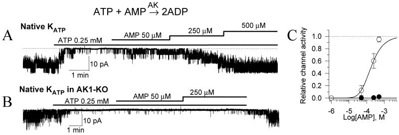
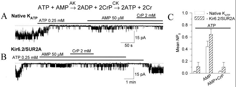
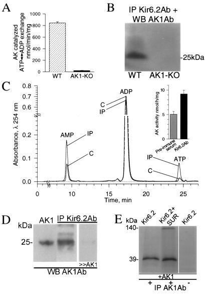
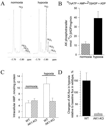
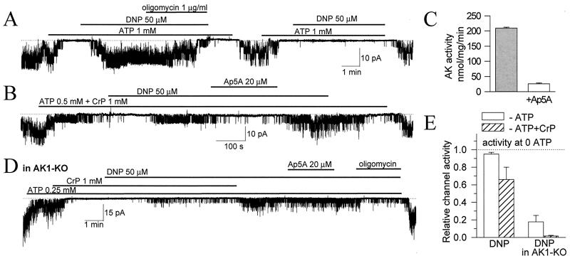
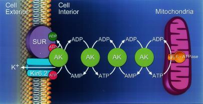
Transduction of energetic signals into membrane electrical events governs vital cellular functions, ranging from hormone secretion and cytoprotection to appetite control and hair growth. Central to the regulation of such diverse cellular processes are the metabolism sensing ATP-sensitive K+ (K(ATP)) channels. However, the mechanism that communicates metabolic signals and integrates cellular energetics with K(ATP) channel-dependent membrane excitability remains elusive. Here, we identify that the response of K(ATP) channels to metabolic challenge is regulated by adenylate kinase phosphotransfer. Adenylate kinase associates with the K(ATP) channel complex, anchoring cellular phosphotransfer networks and facilitating delivery of mitochondrial signals to the membrane environment. Deletion of the adenylate kinase gene compromised nucleotide exchange at the channel site and impeded communication between mitochondria and K(ATP) channels, rendering cellular metabolic sensing defective. Assigning a signal processing role to adenylate kinase identifies a phosphorelay mechanism essential for efficient coupling of cellular energetics with K(ATP) channels and associated functions.


Figures

Figure 1
Figure 1
AMP-dependent regulation of KATP channels is lost in AK1-KO cardiac cells. (A) Recording of native cardiac KATP channels demonstrate activation of ATP-inhibited channels following application of AMP, a pivotal AK substrate. (B) Absence of the AMP effect in cardiomyocytes from AK1-KO mice. (C) Concentration dependence of AMP-induced activation on 250 μM ATP-inhibited KATP channels in control (○) and AK1-KO (●) cardiomyocytes.
Figure 2
Figure 2
CK regulates AK-mediated KATP channel activation. Although AMP activated both native cardiac (A) and recombinant Kir6.2/SUR2A (B) KATP channels, CrP, which promotes ADP scavenging by CK, antagonized AMP-induced KATP channel activation. (C) Average KATP channel activity (mean ± SE) in cardiomyocytes (n = 4; □) and in COS-7 cells expressing recombinant KATP channels (Kir6.2/SUR2A, n = 4; ▨), in the presence of 250 μM ATP, following addition of 50 μM AMP or following addition of AMP plus 2 mM CrP.
Figure 3
Figure 3
AK associates with sarcolemmal KATP channels. (A) Vigorous AK ATP ↔ ADP exchange activity in sarcolemma from wild-type (WT, n = 4; ▨), but not AK1-KO (n = 4; ■) mice. (B) Association of KATP channel protein with AK1 in wild-type, but not AK1-KO. Sarcolemmal KATP channels were immunoprecipitated (IP) with the anti-Kir6.2 Ab (Kir6.2Ab) and probed for AK1 by Western blot (WB) with the anti-AK1 Ab (AK1Ab). (C) HPLC chromatogram shows increased AK activity in KATP channel immunoprecipitates (IP) compared with control (C). The anti-Kir6.2 Ab was used to immunoprecipitate the channel complex from cardiac sarcolemma, while preimmune serum served as control. (D) Anti-AK1 Ab recognized, by Western blot, recombinant AK1 (left lane) and a protein of identical molecular mass in sarcolemmal Kir6.2 immunoprecipitates (center lane), which was competed off by excess of purified AK1 (right lane). (E) Channel proteins were in vitro translated and labeled with [35S]methionine. In the presence of nonlabeled recombinant AK1, the anti-AK1 Ab immunoprecipitated Kir6.2 (left lane) or Kir6.2/SUR subunits (center lane). Channel proteins were not immunoprecipitated by a control Ab (right lane). Proteins were run on denaturing SDS/PAGE, and visualized by Western blotting (B and D) or autoradiography (E).
Figure 4
Figure 4
AK phosphotransfer under metabolic stress. (A) 31P NMR spectra of guinea pig heart extracts under normoxic and hypoxic conditions. Incorporation of 18O into β-phosphoryls of ADP, which reflects the rate of AK-catalyzed phosphotransfer, induces the appearance of three 18O-labeled species (18O1, 18O2, and 18O3). Incorporation of 18O was significantly higher under hypoxia. (B) Average values for AK-catalyzed phosphotransfer flux in normoxia (n = 5; ▩) and hypoxia (n = 5; ■) in guinea pig heart determined by 18O/31P NMR spectroscopy. (C) AMP levels in wild-type (WT, n = 5; □) and AK1-KO (n = 5; ▨) mouse heart under normoxia and hypoxia determined by HPLC. (D) Blunted AK phosphotransfer response in the AK1-KO (n = 5; ░⃞) compared with the wild-type (n = 5; ■) mouse heart in hypoxia.
Figure 5
Figure 5
AK communicates mitochondrial signals to KATP channels. (A) The mitochondrial uncoupler, DNP, induced vigorous KATP channel activation which was irreversibly inhibited by oligomycin, a mitochondrial FoF1-ATPase antagonist (n = 5). (B) In the presence of CrP, DNP produced lower activation of KATP channels as part of mitochondria-generated ADP was scavenged by the CrP/CK system. Ap5A, which at 20 μM inhibited AK activity in cardiac sarcolemma (n = 3; C), suppressed AK-mediated channel activation (n = 5). (D) In AK1-KO cardiomyocytes, KATP channel activation by DNP was blunted and insensitive to Ap5A, but inhibited by oligomycin (n = 3). (E) Average (mean ± SE) DNP-induced KATP channel activity in wild-type (left) and AK1-KO (right) cardiomyocytes in 0.5 mM ATP (□) or 0.5 mM ATP plus 1 mM CrP (▨). Bars were constructed from three to five recordings for each data point.
Figure 6
Figure 6
AK phosphotransfer communicates mitochondria-generated signals to KATP channels. AK molecules form a phosphorelay network connecting mitochondria with the cell membrane. Under hypoxic stress, mitochondrial FoF1-ATPase consumes cellular ATP generating ADP, which is delivered to the KATP channel through the chain of sequential AK-catalyzed phosphotransfer reactions. The inwardly rectifying potassium channel (Kir6.2) and the SUR are the pore-forming and regulatory subunits of the KATP channel complex.

References

    1. Weiss J N, Lamp S T. Science. 1987;238:67–69. - PubMed
    1. O'Rourke B, Ramza B M, Marban E. Science. 1994;265:962–966. - PubMed
    1. Noma A. Nature (London) 1983;305:147–148. - PubMed
    1. Ashcroft F M, Harrison D E, Ashcroft S J. Nature (London) 1984;312:446–448. - PubMed
    1. Inagaki N, Gonoi T, Clement J P, Namba N, Inazawa J, Gonzalez G, Aguilar-Bryan L, Seino S, Bryan J. Science. 1995;270:1166–1170. - PubMed

Publication types

MeSH terms

LinkOut - more resources