. 2001 Jun 19;98(13):7593-8.
doi: 10.1073/pnas.121179898. Epub 2001 Jun 5.

Effects of anesthesia on functional activation of cerebral blood flow and metabolism

Affiliations

Effects of anesthesia on functional activation of cerebral blood flow and metabolism

Y Nakao et al. Proc Natl Acad Sci U S A. .

Abstract

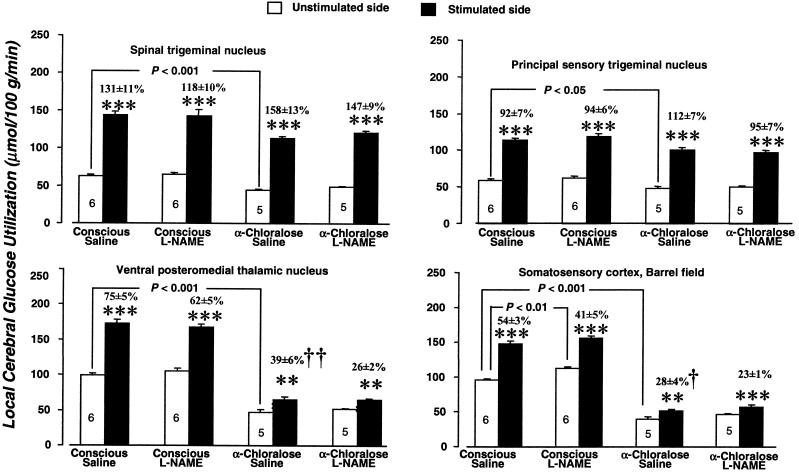
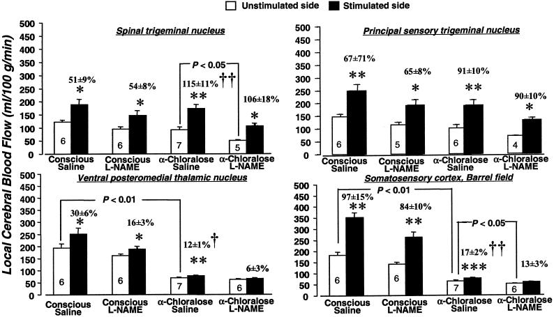
Functional brain mapping based on changes in local cerebral blood flow (lCBF) or glucose utilization (lCMR(glc)) induced by functional activation is generally carried out in animals under anesthesia, usually alpha-chloralose because of its lesser effects on cardiovascular, respiratory, and reflex functions. Results of studies on the role of nitric oxide (NO) in the mechanism of functional activation of lCBF have differed in unanesthetized and anesthetized animals. NO synthase inhibition markedly attenuates or eliminates the lCBF responses in anesthetized animals but not in unanesthetized animals. The present study examines in conscious rats and rats anesthetized with alpha-chloralose the effects of vibrissal stimulation on lCMR(glc) and lCBF in the whisker-to-barrel cortex pathway and on the effects of NO synthase inhibition with N(G)-nitro-L-arginine methyl ester (L-NAME) on the magnitude of the responses. Anesthesia markedly reduced the lCBF and lCMR(glc) responses in the ventral posteromedial thalamic nucleus and barrel cortex but not in the spinal and principal trigeminal nuclei. L-NAME did not alter the lCBF responses in any of the structures of the pathway in the unanesthetized rats and also not in the trigeminal nuclei of the anesthetized rats. In the thalamus and sensory cortex of the anesthetized rats, where the lCBF responses to stimulation had already been drastically diminished by the anesthesia, L-NAME treatment resulted in loss of statistically significant activation of lCBF by vibrissal stimulation. These results indicate that NO does not mediate functional activation of lCBF under physiological conditions.


Figures

Figure 1
Figure 1
Effects of α-chloralose anesthesia and L-NAME (30 mg/kg) on baseline rates of glucose utilization and on the increases in glucose utilization due to unilateral vibrissal stimulation in four stations of the whisker-to-barrel cortex pathway. Bar heights and error bars represent mean rates of local glucose utilization + SEM obtained in the number of rats indicated. **, P < 0.01; ***, P < 0.001 for side-to-side differences between stimulated and unstimulated sides (paired t test with Bonferroni correction). Bracketed P values are for comparison of the values of glucose utilization in the structures and sides indicated by the brackets (unpaired t test with Bonferroni correction). †, P < 0.05; ††, P < 0.01, statistical significance of difference in percent increase in glucose utilization attributable to stimulation in indicated condition from percent increases due to stimulation found in conscious saline-treated controls, determined by Kruskal–Wallis test (one-way ANOVA by ranks) with Dunn's test.
Figure 2
Figure 2
Effects of α-chloralose anesthesia and L-NAME (30 mg/kg) on baseline blood flow and increases in blood flow due to vibrissal stimulation in four stations of the whisker-to-barrel cortex pathway. Bar heights and error bars represent mean rates of blood flow + SEM obtained in the number of rats indicated. *, P < 0.05; **, P < 0.01; ***, P < 0.001 for side-to-side differences between stimulated and unstimulated sides (paired t test with Bonferroni correction). Bracketed P values are for comparison of the values of blood flow in the structures and sides indicated by the brackets (unpaired t test with Bonferroni correction). †, P < 0.05; ††, P < 0.01, statistical significance of difference in percent increase in blood flow attributable to stimulation in indicated condition from percent increases due to stimulation found in conscious saline-treated controls, determined by Kruskal–Wallis test (one-way ANOVA by ranks) with Dunn's test.

References

    1. Kadekaro M, Crane A M, Sokoloff L. Proc Natl Acad Sci USA. 1985;82:6010–6013. - PMC - PubMed
    1. Sokoloff L. Neurochem Res. 1999;24:321–329. - PubMed
    1. Mata M, Fink D J, Gainer H, Smith C B, Davidsen L, Savaki H, Schwartz W J, Sokoloff L. J Neurochem. 1980;34:213–215. - PubMed
    1. Pellerin L, Magistretti P J. Proc Natl Acad Sci USA. 1994;91:10625–10629. - PMC - PubMed
    1. Takahashi S, Driscoll B F, Law M J, Sokoloff L. Proc Natl Acad Sci USA. 1995;92:4616–4620. - PMC - PubMed

MeSH terms

LinkOut - more resources