. 2001 Jun 19;98(13):7414-9.
doi: 10.1073/pnas.131090498. Epub 2001 Jun 5.

A gene family required for human germ cell development evolved from an ancient meiotic gene conserved in metazoans

Affiliations

A gene family required for human germ cell development evolved from an ancient meiotic gene conserved in metazoans

E Y Xu et al. Proc Natl Acad Sci U S A. .

Abstract

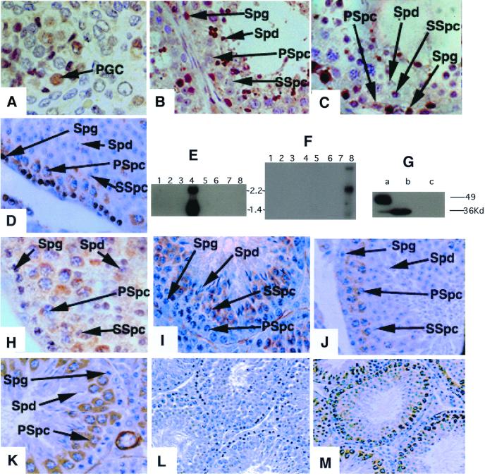
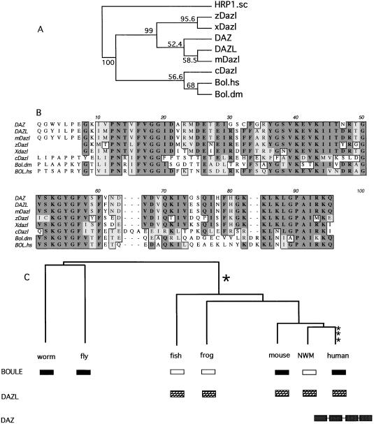
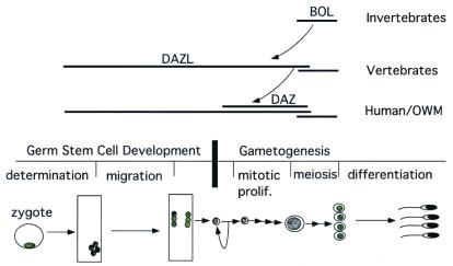
The Deleted in AZoospermia (DAZ) genes encode potential RNA-binding proteins that are expressed exclusively in prenatal and postnatal germ cells and are strong candidates for human fertility factors. Here we report the identification of an additional member of the DAZ gene family, which we have called BOULE. With the identification of this gene, it is clear that the human DAZ gene family contains at least three members: DAZ, a Y-chromosome gene cluster that arose 30-40 million years ago and whose deletion is linked to infertility in men; DAZL, the "father" of DAZ, a gene that maps to human chromosome 3 and has homologs required for both female and male germ cell development in other organisms; and BOULE, a gene that we propose is the "grandfather" of DAZ and maps to human chromosome 2. Human and mouse BOULE resemble the invertebrate meiotic regulator Boule, the proposed ortholog of DAZ, in sequence and expression pattern and hence likely perform a similar meiotic function. In contrast, the previously identified human DAZ and DAZL are expressed much earlier than BOULE in prenatal germ stem cells and spermatogonia; DAZL also is expressed in female germ cells. These data suggest that homologs of the DAZ gene family can be grouped into two subfamilies (BOULE and DAZL) and that members of the DAZ family evolved from an ancestral meiotic regulator, Boule, to assume distinct, yet overlapping, functions in germ cell development.


Figures

Figure 1
Figure 1
Human BOULE (BOULE) encodes a protein that is homologous to fly Boule and human DAZ/DAZL. (A) Diagram of BOULE, DAZL, and DAZ proteins. Black boxes represent the RNA-binding domains, hatched boxes represent the DAZ repeats. (B) Alignment of RNA-binding domains of fly Boule, human BOULE, human DAZL, and human DAZ proteins. * indicates conserved residues, and • indicates similar residues among all four proteins. Shadowed boxes outline amino acids shared between Boule and BOULE but different from DAZ or DAZL. Open boxes indicate amino acids shared between DAZ or DAZL and BOULE but not with fly Boule. Solid arrows indicate shared splice sites and open arrows indicate unique splice sites in Boule and/or BOULE. Dashed arrow indicates a shared splice site if we consider the two amino acids (positions 73 and 74) as a single addition/deletion. (C) Conservation of the DAZ repeats in DAZ and BOULE homologs. Shade indicates identical or similar amino acids.
Figure 2
Figure 2
BOULE expression is distinct from that of DAZ and DAZL. DAZ is expressed in prenatal primordial germ cells, spermatogonial stem cells, and spermatocytes. DAZL is expressed in both the male and female germ line. BOULE is expressed in the cytoplasm of pachytene spermatocytes, persists through meiosis, and decreases in early spermatids. (A) DAZ staining of fetal primordial germ cells of the human testes; similar staining is observed in female primordial germ cells (6, 18). (B) Human testes staining with antisera that recognize DAZL only; DAZL is expressed in spermatogonia, early and late spermatocytes, and postmeiotic cells. Staining of human ovary with this antisera indicate cytoplasmic staining of oocytes (6, 18). (C) Human testis section stained using antisera that recognize DAZ only. DAZ is expressed in spermatogonia and early spermatocytes, but is absent from late spermatocytes or postmeiotic cells. (D) Mouse Dazl is present in spermatogonia and early and late spermatocytes as in human testes. As in humans, female mice also express Dazl in the germ cells (6, 12). (E) A Northern blot with polyadenylated RNA from different human tissues. Blot was hybridized with human BOULE cDNA that detects two testes specific transcripts. Lanes: 1, spleen; 2, thymus; 3, prostate; 4, testis; 5, ovary; 6, small intestine; 7, colon; and 8, leukocyte. (F) A Northern blot with polyadenylated RNA from mouse tissues. Blot was hybridized with human BOULE cDNA that detects three testes-specific transcripts. Lanes: 1, heart; 2, brain; 3, spleen; 4, lung; 5, liver; 6, muscle; 7, kidney; and 8, testis. (G) Anti-BOULE antisera detects a single 32-kDa protein in mouse testes (b) and a similar size protein in human testes (a) but not in other human or mouse tissues (data not shown), nor does it recognize DAZ protein expressed in yeast strain, RRY618 (c). The 50-kDa band in human testes is nonspecific as it is detected by preimmune also. (H) BOULE staining in human testis section is also restricted to cytoplasm of spermatocytes; no staining of spermatogonial stem cells is observed. (I) Stage III seminiferous tubules. BOULE is expressed in round spermatids (Spd) and secondary spermatocytes but not in spermatogonia (Spg) or primary spermatocytes (Spc). (J) Stage VII seminiferous tubule. BOULE is expressed in the cytoplasm of pachytene spermatocytes. There is no staining in spermatogonia and spermatids. (K) Stage X–XI seminiferous tubules. BOULE expression peaks in late pachytene stage spermatocytes. (L and M) Lower-magnification view of staining with preimmune and anti-BOULE antisera. (L) Preimmune of BOULE antisera (×200). (M) BOULE antisera (×200). Spg, spermatogonial cells; Spc, spermatocytes; SSpc, secondary spermatocytes; Spd, spermatids; Pgc, primordial germ cell. Unless noted, all pictures were taken at the same magnification (×630). DAZ and DAZL antisera are described (6, 18).
Figure 3
Figure 3
DAZ/DAZL evolved from Boule. (A) Phylogenetic tree based on the RNA-binding domains of human BOULE (BOULE.hs), fly Boule (BOULE.dm), human DAZL (DAZL), and known homologs from mouse (mDazl), worm (cDazl), zebrafish (zDazl), and frog (Xdazl). Yeast HRP1 (HRP1.sc), which belongs to the same RNP family as DAZ/BOULE, was used as an outgroup. Both maximum parsimony and distance-based methods using PHYLIP 3.5 (J. Felsenstein) produce trees of similar topology. The consensus tree presented here is built with maximum parsimony method (PROTPARS). Numbers indicate the percentage of the bootstrapping trials in which an identical node was produced. The number of bootstrap replicates was 100. The position of DAZ was placed outside its ancestor mouse Dazl due to greater divergence in DAZ than DAZL. (B) Multiple sequence alignment of RNP domains in BOULE and DAZL homologs. Conserved residues are boxed (dark shadow is identical and light shadow is similar). (C) Model of evolutionary history of BOULE/DAZ family. Dark box represents BOULE homologs and hatched box represents DAZL homologs. Open box indicates the inferred presence of a BOULE homolog that has yet to be identified. The DAZ gene cluster on the Y chromosome is represented by four hatched boxes linked together to indicate the presence of at least four genes in tandem (3, 25). Vertical hatch box indicates the probable period of gene duplication. NWM, New World monkey.
Figure 4
Figure 4
The ancient meiotic regulator, Boule, is conserved in all metazoans and gave rise to a gene family required for novel vertebrate germ cell functions. Expression pattern of each member is indicated by extent of each horizontal line. Meiotic expression of BOULE is seen in fly, mice and humans (primary spermatocytes) and in female oocytes before meiosis (8, 11). This meiotic function is likely conserved throughout metazoans. DAZL evolved a novel function required for germ stem cell proliferation or differentiation that is unique to vertebrates. DAZL is expressed in multiple compartments from germ stem cells to mature spermatocytes. DAZ arose from DAZL recently in the primate lineage and is expressed in multiple compartments from germ stem cells to meiotic cells. OWM, Old World monkey. Major stages of germ cell development are diagramed in the middle to provide a timeline for DAZ family gene expression. The curved arrow indicates a duplication.

Comment in

  • Rolling back to BOULE.
    Haag ES. Haag ES. Proc Natl Acad Sci U S A. 2001 Jun 19;98(13):6983-5. doi: 10.1073/pnas.141237898. Proc Natl Acad Sci U S A. 2001. PMID: 11416173 Free PMC article. Review. No abstract available.

References

    1. Reijo R, Lee T Y, Salo P, Alagappan R, Brown L G, Rosenberg M, Rozen S, Jaffe T, Straus D, Hovatta O, et al. Nat Genet. 1995;10:383–393. - PubMed
    1. Cooke H J, Elliot D J. Trends Genet. 1997;13:87–92. - PubMed
    1. Saxena R, Brown L G, Hawkins T, Alagappan R K, Skaletsky H, Reeve M P, Reijo R, Rozen S, Dinulos M B, Disteche C M, Page D C. Nat Genet. 1996;14:292–299. - PubMed
    1. Seboun E, Barbaux S, Bourgeron T, Nishi S, Algonik A, Egashira M, Nikkawa N, Bishop C, Fellow M, McElreavey K, Kasahara M. Genomics. 1997;41:227–235. - PubMed
    1. Yen P H, Chai N N, Salido E C. Hum Mol Genet. 1996;5:2013–2017. - PubMed

Publication types

MeSH terms

Associated data