. 2001 Jul 3;98(14):8133-8.
doi: 10.1073/pnas.131186798. Epub 2001 Jun 5.

The role of haustoria in sugar supply during infection of broad bean by the rust fungus Uromyces fabae

Affiliations

The role of haustoria in sugar supply during infection of broad bean by the rust fungus Uromyces fabae

R T Voegele et al. Proc Natl Acad Sci U S A. .

Abstract

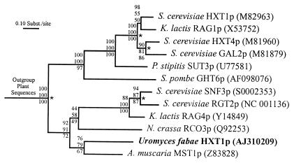
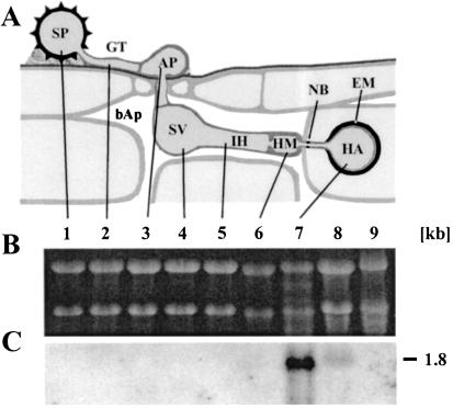
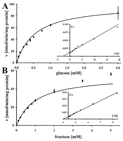
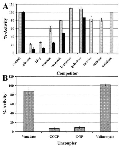
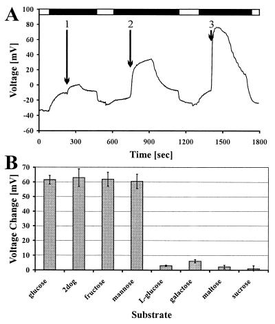
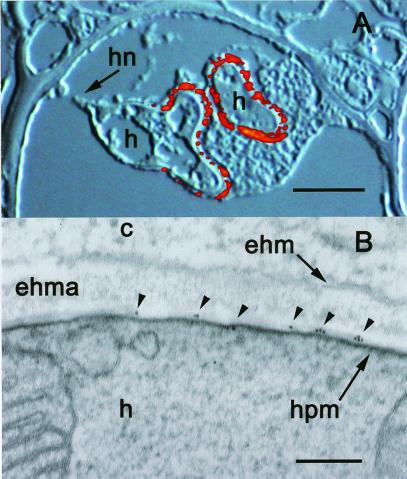
Biotrophic plant pathogenic fungi differentiate specialized infection structures within the living cells of their host plants. These haustoria have been linked to nutrient uptake ever since their discovery. We have for the first time to our knowledge shown that the flow of sugars from the host Vicia faba to the rust fungus Uromyces fabae seems to occur largely through the haustorial complex. One of the most abundantly expressed genes in rust haustoria, the expression of which is negligible in other fungal structures, codes for a hexose transporter. Functional expression of the gene termed HXT1 in Saccharomyces cerevisiae and Xenopus laevis oocytes assigned a substrate specificity for D-glucose and D-fructose and indicated a proton symport mechanism. Abs against HXT1p exclusively labeled haustoria in immunofluorescence microscopy and the haustorial plasma membrane in electron microscopy. These results suggest that the fungus concentrates this transporter in haustoria to take advantage of a specialized compartment of the haustorial complex. The extrahaustorial matrix, delimited by the plasma membranes of both host and parasite, constitutes a newly formed apoplastic compartment with qualities distinct from those of the bulk apoplast. This organization might facilitate the competition of the parasite with natural sink organs of the host.


Figures

Figure 1
Figure 1
Homology of HXT1p to other fungal hexose transporters. Shown is the maximum likelihood tree. Distance-based neighbor-joining and maximum-parsimony methods produced similar topologies. Bootstrap values for maximum likelihood, distance-based neighbor-joining, and maximum-parsimony methods (from top to bottom) are shown at the corresponding branch points. Sequences are identified by species and transporter name. Accession numbers are given in parentheses. The asterisks mark gene-duplication events.
Figure 2
Figure 2
HXT1 is a single-copy gene. Total DNA of U. fabae was prepared from germinated spores and digested with EcoRI (lane 1), BamHI (lane 2), EcoRV (lane 3), PvuII (lane 4), and PstI (lane 5). Southern blot analysis, using an HXT1-specific cDNA probe, produced single bands in all cases but PstI. For PstI, two bands were obtained because of an internal PstI site at sequence position 4,786. Numbers on the right give the size of the molecular weight marker in kb.
Figure 3
Figure 3
HXT1 transcripts are found only in haustoria and infected leaves. (A) Schematic representation of rust infection structures. (B) Ethidium bromide-stained denaturing agarose gel (loading control). (C) Northern blot of the gel depicted in B. Lane 1, uredospore (SP); and lane 2, germtube (GT) after 4-h germination. Lanes 3–6, in vitro infection structures harvested at the following stages: 3, appressorium (AP) stage (6-h); 4, substomatal vesicle (SV) stage (12-h); 5, infection hyphae (IH) stage (18-h); 6, haustorial mother cell (HM) stage (21-h). Lane 7, isolated haustoria (HA); lane 8, infected leaves; and lane 9, noninfected leaves. bAp, bulk apoplast; NB, neckband; EM, extrahaustorial matrix. The number on the right gives the size estimate in kb.
Figure 4
Figure 4
HXT1p kinetics with D-glucose (A) or D-fructose (B) as substrate. Michaelis–Menten plots with corresponding Lineweaver–Burke plots (Insets). Radio-labeled sugars were used in the yeast system to obtain transport rates of HXT1p.
Figure 5
Figure 5
Competition and uncoupling experiments. (A) Competition experiments using 300 μM D-glucose (gray bars) or 800 mM D-fructose (black bars) and competitor in 10-fold excess in the yeast system. Activity is reported in % of the control without competitor added. The respective control rates were 22.6 ± 6.5 nmol/min/mg of protein (glucose) and 10 ± 0.2 nmol/min/mg of protein (fructose). (B) Uncoupling experiments with 300 μM D-glucose as substrate in the yeast system. Activity is reported in % with respect to the activity obtained by using the respective solvent. The respective control rate (no addition) was 18.7 ± 1.1 nmol/min/mg of protein.
Figure 6
Figure 6
Electrophysiological data obtained with the oocyte system. (A) Voltage trace showing the depolarization of the membrane in response to the addition of D-glucose (1:0.1 mM, 2:1 mM, and 3:10 mM). Black bars indicate use of pH 5.0 to create a proton gradient, white bars refer to use of pH 7.5. (B) Membrane depolarization in response to different sugars. Test substances were used at a concentration of 5 mM after pH downshift.
Figure 7
Figure 7
Localization of HXT1p in the periphery of fully developed haustoria and along the haustorial plasma membrane. (A) Superimposed Nomarski differential interference contrast and fluorescence images depicting two haustoria. Labeling of HXT1p with S651p resulted only in fluorescence signals in the periphery of the distal parts of the haustorium; proximal parts and haustorial neck are not labeled (visible on the left haustorium). h, haustorium; hn, haustorial neck. (Bar, 5 μm.) (B) Electron micrograph depicting considerable gold labeling along the haustorial plasma membrane (hpm) only (small arrows), but no labeling over the h, the extra haustorial matrix (ehma), the extrahaustorial membrane (ehm), or the plant cytoplasm (c). (Bar, 0.1 μm.)

Comment in

References

    1. Heath M C, Skalamera D. Adv Bot Res. 1997;24:195–225.
    1. Bushnell W R. Annu Rev Phytopathol. 1972;10:151–176.
    1. Mendgen K, Struck C, Voegele R T, Hahn M. Physiol Mol Plant Pathol. 2000;56:141–145.
    1. de Bary A. Vergleichende Morphologie der Pilze, Mycetozoen und Bacterien. Leipzig, Germany: Engelmann; 1884.
    1. Hahn M, Mendgen K. Protoplasma. 1992;170:95–103.

Publication types

Substances

Associated data