. 2001 Jun 5;98(12):6836-41.
doi: 10.1073/pnas.111057298.

The copper transporter CTR1 provides an essential function in mammalian embryonic development

Affiliations

The copper transporter CTR1 provides an essential function in mammalian embryonic development

Y M Kuo et al. Proc Natl Acad Sci U S A. .

Abstract

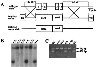
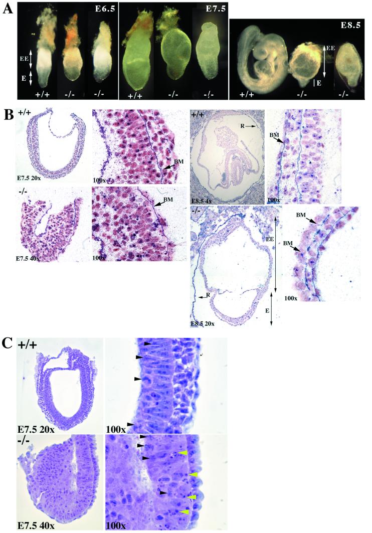
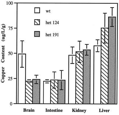
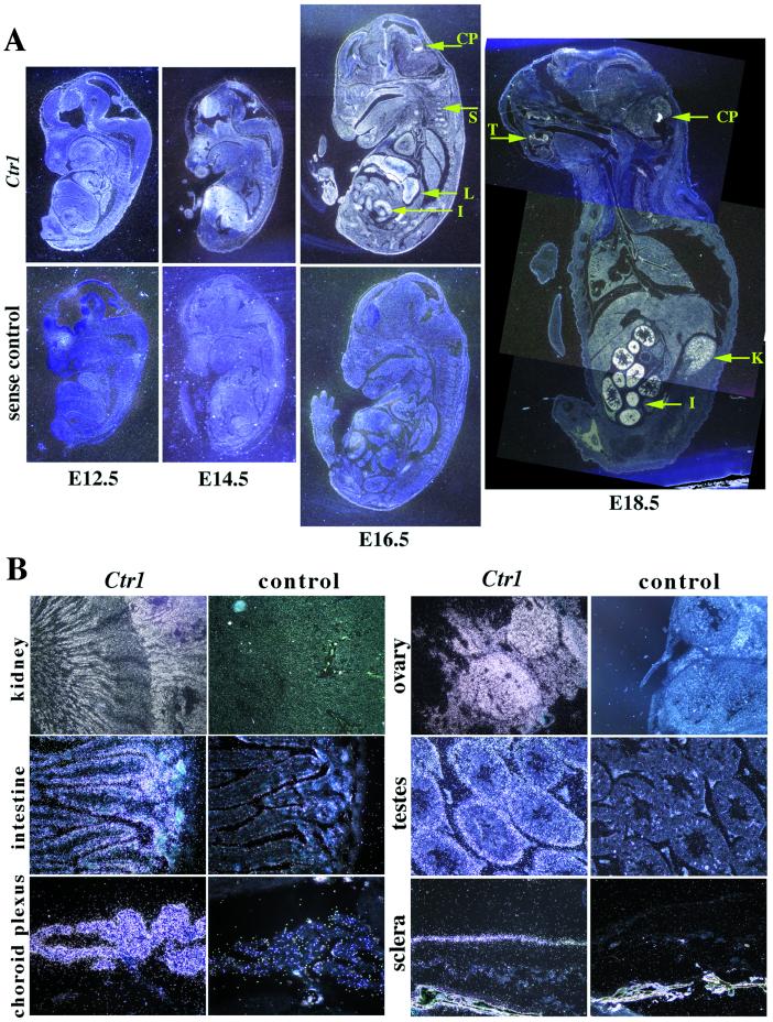
Copper serves as an essential cofactor for a variety of proteins in all living organisms. Previously, we described a human gene (CTR1;SLC31A1) that encodes a high-affinity copper-uptake protein and hypothesized that this protein is required for copper delivery to mammalian cells. Here, we test this hypothesis by inactivating the Ctr1 gene in mice by targeted mutagenesis. We observe early embryonic lethality in homozygous mutant embryos and a deficiency in copper uptake in the brains of heterozygous animals. Ctr1(-/-) embryos can be recovered at E8.5 but are severely developmentally retarded and morphologically abnormal. Histological analysis reveals discontinuities and variable thickness in the basement membrane of the embryonic region and an imperfect Reichert's membrane, features that are likely due to lack of activity in the collagen cross-linking cupro-enzyme lysyl oxidase. A collapsed embryonic cavity, the absence of an allantois, retarded mesodermal migration, and increased cell death are also apparent. In the brains of heterozygous adult mice, which at 16 months are phenotypically normal, copper is reduced to approximately half compared with control littermates, implicating CTR1 as the required port for copper entry into at least this organ. A study of the spatial and temporal expression pattern of Ctr1 during mouse development and adulthood further shows that CTR1 is ubiquitously transcribed with highest expression observed in the specialized epithelia of the choroid plexus and renal tubules and in connective tissues of the eye, ovary, and testes. We conclude that CTR1 is the primary avenue for copper uptake in mammalian cells.


Figures

Figure 1
Figure 1
Targeted disruption of Ctr1. (A) Schematic illustration of the Ctr1 gene, the targeting construct, and the predicted mutant allele. The four exons of Ctr1 are shown. Homologous recombination in the regions indicated X should result in the predicted mutant allele. (B) Southern blot verification of correctly targeted ES cell clones. Hybridization of SpeI-digested DNA from ES lines probed with a radiolabeled 3′ external probe reveals a targeting event as a pair of fragments. (C) PCR analysis of F1 heterozygous mouse genomic DNA using two pairs of primers, as described under Materials and Methods. The 730-bp fragment corresponds to the normal gene, and the 421-bp fragment is derived from the correctly targeted gene.
Figure 2
Figure 2
Phenotypic analysis of wild-type and mutant embryos. (A) Whole-mount gross morphology of wild-type and Ctr1−/− embryos at E6.5, E7.5, and E8.5. The embryos are oriented such that the embryonic region (E) is located at the bottom and the extra-embryonic structures are on the top (EE). All embryos were genotyped by PCR after photography. Identical phenotypes were observed in both the 124 and 191 lines, and wild-type embryos were indistinguishable from heterozygotes. Photographs were taken on a Nikon SMZ 800 dissecting microscope. E6.5 embryos were photographed at ×6.5, E7.5 at ×5, and E8.5 at ×4 with a ×2.5 projection lens. (B) Histological examination of sectioned embryos. E7.5 and E8.5 wild-type and mutant were stained with anti-collagen IV staining and counterstained with Fast Red. At E7.5, the basement membrane (BM) of the mutant embryo appears intact. At E8.5, the mutant extra-embryonic region (EE) and underdeveloped embryonic region (E) are seen. Note the Reichert's membrane (R) is broken in the E8.5 mutants but intact in the wild-type E8.5. At ×100, the basement membrane is intact in the wild type, but gaps along the basement membrane are apparent in the mutant, as indicated by the arrows. (C) Hematoxylin and eosin staining of sagittal sections of E7.5 wild-type and mutant embryos. Only the embryonic region is shown because the upper extra-embryonic region was removed for genotyping by PCR. At ×100 in the mutant, areas of apoptosis can be seen (indicated by yellow arrowheads); however, mitosis is unaffected (indicated by black arrowheads). In the wild-type normal section at ×100, mitosis can be seen as indicated by the black arrowheads.
Figure 3
Figure 3
Copper content of brain, small intestine, kidney, and liver from heterozygous and wild-type 6-week-old male littermates. Six heterozygous mice from each independent line 124 and 191 and four wild-type littermate mice were analyzed. Digested tissue samples were assayed three times as normalized to wet weight of tissue. The measurements were averaged, and the standard deviation for each was determined by using the MICROSOFT EXCEL program.
Figure 4
Figure 4
In situ hybridization of Ctr1 in wild-type embryos and adult tissues. (A) Temporal and spatial expression of Ctr1 in the developing mouse. At E14.5, expression can be observed in the forebrain, the liver, the nasal regions, and the somites. By E16.5, Ctr1 expression is ubiquitous, with high expression in the liver (L), intestine (I), somites (S), and choroid plexus (CP). By E18.5 expression is more restricted to the choroid plexus (CP), kidney (K), intestine (I), and tooth buds (T). (B) Expression of Ctr1 in adult tissues. Ctr1 is expressed in the tubules in the inner cortex and inner medulla of the kidney, the villi of the small intestine, the choroid plexus of the brain, the stroma of the ovary, the seminiferous tubules of the testes, and the sclera of the eye. Sections were photographed under dark-field illumination. The E12.5 embryo was photographed at ×4, the E14.5, at ×2, and the E16.5 and E18.5 at ×1. The kidney was photographed at ×4, the ovary, testes, and eye at ×20, and the intestine and choroid plexus at ×40.

Comment in

  • Mining copper transport genes.
    Andrews NC. Andrews NC. Proc Natl Acad Sci U S A. 2001 Jun 5;98(12):6543-5. doi: 10.1073/pnas.131192498. Proc Natl Acad Sci U S A. 2001. PMID: 11390990 Free PMC article. No abstract available.

References

    1. Harris E D. Proc Soc Exp Biol Med. 1991;196:130–140. - PubMed
    1. Bull P C, Cox D W. Trends Genet. 1994;10:246–252. - PubMed
    1. Danks D M. In: The Metabolic Basis of Inherited Disease. Scriver C R, Beaudet A L, Sly W S, Valle D, editors. Vol. 2. New York: McGraw–Hill; 1995. pp. 2211–2235.
    1. Vulpe C, Packman S. Annu Rev Nutr. 1995;15:293–322. - PubMed
    1. Vulpe C, Levinson B, Whitney S, Packman S, Gitschier J. Nat Genet. 1993;3:7–13. - PubMed

Publication types