. 2001 Jun 5;98(12):6848-52.
doi: 10.1073/pnas.111058498.

The metallochaperone Atox1 plays a critical role in perinatal copper homeostasis

Affiliations

The metallochaperone Atox1 plays a critical role in perinatal copper homeostasis

I Hamza et al. Proc Natl Acad Sci U S A. .

Abstract

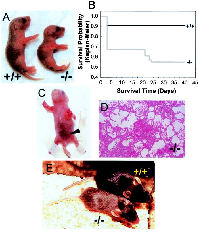
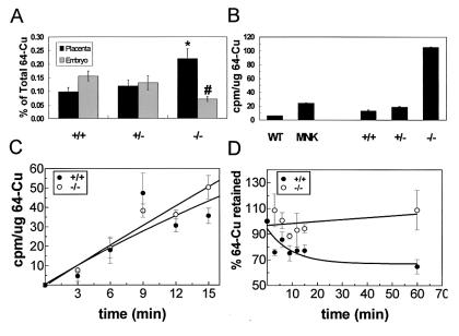
Copper plays a fundamental role in the biochemistry of all aerobic organisms. The delivery of this metal to specific intracellular targets is mediated by metallochaperones. To elucidate the role of the metallochaperone Atox1, we analyzed mice with a disruption of the Atox1 locus. Atox1(-/-) mice failed to thrive immediately after birth, with 45% of pups dying before weaning. Surviving animals exhibited growth failure, skin laxity, hypopigmentation, and seizures because of perinatal copper deficiency. Maternal Atox1 deficiency markedly increased the severity of Atox1(-/-) phenotype, resulting in increased perinatal mortality as well as severe growth retardation and congenital malformations among surviving Atox1(-/-) progeny. Furthermore, Atox1-deficient cells accumulated high levels of intracellular copper, and metabolic studies indicated that this defect was because of impaired cellular copper efflux. Taken together, these data reveal a direct role for Atox1 in trafficking of intracellular copper to the secretory pathway of mammalian cells and demonstrate that this metallochaperone plays a critical role in perinatal copper homeostasis.


Figures

Figure 1
Figure 1
Generation of the Atox1 null allele. (A) The genomic locus of the mouse Atox1 gene disrupted by the promoterless β-galactosidase-neomycin (β-geo) cassette. The relevant restriction sites, location of the β-geo insertion (arrowhead) between exons 1 and 2, the 5′ probe for Southern analysis (horizontal bar), and location of sequences used as primer sets for genotyping (horizontal arrows) are indicated (En2-SA, engrailed 2 splice acceptor site; IRES, internal ribosome entry site). (B) Southern blot analysis of genomic DNA digested with EcoRI from Atox1+/+, Atox1+/−, and Atox1−/− embryos and probed with the 5′ probe. (C) RNA blot analysis of 10 μg of total RNA isolated from E12.5 embryonic fibroblasts and probed with the Atox1 or 18S (Ambion) DNA probe. (D) Immunoblot analysis of Atox1 protein in E12.5 embryonic fibroblasts. One-hundred-microgram cell lysates were separated by 10–20% Tricine SDS/PAGE gel and probed with ATOX1 antisera followed by chemiluminescent detection.
Figure 2
Figure 2
Atox1 deficiency results in growth failure and mortality. (A) Images of Atox1+/+ (Left) and Atox1−/− pups (Right) depicting growth differences at P3. (B) Kaplan–Meier plot of survival in progeny from Atox1+/− ♂ × Atox1+/− ♀ mating (n = 166). Note initial drop by P3, followed by another decrease because of seizures around weaning age. Abdominal hemorrhage (arrowhead) (C) and micrograph (100×) depicting pulmonary alveolar hemorrhage (D) observed in some Atox1−/− newborn pups. (E) Twenty-three-day-old weanling Atox1+/+ (Right) and Atox1−/− (Left) mice. Note size difference and the hypopigmentation of tail and coat color.
Figure 3
Figure 3
Atox1 is essential for normal copper homeostasis. (A) Placental copper transport was determined in E14.5 pregnant females from Atox1+/− ♂ × Atox1+/− ♀ mating. Fifty micrograms of 64Cu was injected intravenously into pregnant dams, and 24 h later the placenta and embryo were dissected from maternal tissue and 64Cu incorporation quantified in a gamma counter. Data represent the mean ± SEM from four separate litters and at least 10 animals of each genotype. Each data point is calculated as the amount of 64Cu retained by the tissue relative to the total amount of 64Cu injected into the mother (*, P < 0.05 among the +/+ and −/− placenta and +/+ and −/− embryos; #, P < 0.01 between −/− placenta and −/− embryos; P values were calculated by using a one-way ANOVA with Student–Newman–Keuls multiple comparison test). (B) 64Cu retention was determined in cultured fibroblasts derived from Atox1 mouse embryos (E12.4 +/+, +/−, and −/−) or mouse cell lines (wt, 802–5; MNK, 802–1). Cells (2 × 10⁵) were incubated with 8 × 10⁶ cpm of 64Cu. After 68 h, the cells were washed, lysed, and analyzed for 64Cu retained by using a gamma counter. Each data point represents the mean ± SEM from three separate experiments performed in triplicate and expressed as cpm/μg of total cell protein. Copper uptake (C) and copper efflux (D) were performed by incubating MEFs with 200 nM 64Cu per 4 × 10⁴ cells. For efflux, cells were pulsed with 64Cu for 1 h followed by several chase periods with unsupplemented culture media. Cells were harvested at time intervals indicated and processed as described above.
Figure 4
Figure 4
Atox1−/− progeny exhibit a maternal effect. (A) Images of Atox1+/− (Left) and Atox1−/− (Right) P8 pups from Atox1+/− ♂ × Atox1−/− ♀ mating. Note skin laxity on the dorsal surface (arrowhead), hypopigmentation, and growth retardation (body weight: +/− = 3.31 g, −/− = 1.74 g). (B) Tyrosinase activity was determined in 100 μg of skin tissue extract separated on a 4–20% SDS/PAGE gel and assayed for dopa oxidase activity. Each lane represents a P8 skin sample from Atox1+/− ♂ × Atox1−/− ♀ progeny. (C) Placental copper transport was determined in Atox1+/− and Atox1−/− pregnant females mated to the indicated males. Data represent the mean ± SEM from three separate litters and at least four animals of each genotype (*, P < 0.01 for −/− placentas and −/− embryos derived from Atox1+/− versus Atox1−/− mothers). (D) Image of Atox1−/− P2 pup showing microphthalmia (Left) and Atox1+/− littermate (Right) from Atox1+/− ♂ × Atox1−/− ♀ mating.

Comment in

  • Mining copper transport genes.
    Andrews NC. Andrews NC. Proc Natl Acad Sci U S A. 2001 Jun 5;98(12):6543-5. doi: 10.1073/pnas.131192498. Proc Natl Acad Sci U S A. 2001. PMID: 11390990 Free PMC article. No abstract available.

References

    1. Pena M M, Lee J, Thiele D J. J Nutr. 1999;129:1251–1260. - PubMed
    1. Culotta V C, Gitlin J D. In: The Metabolic and Molecular Bases of Inherited Diseases. Scriver C R, Beaudet A L, Sly W S, Valle D, editors. Vol. 2. New York: McGraw–Hill; 2001. pp. 3105–3126.
    1. Valentine J, Gralla E B. Science. 1997;278:817–818. - PubMed
    1. O'Halloran T V, Culotta V C. J Biol Chem. 2000;275:25057–25060. - PubMed
    1. Rae T D, Schmidt P J, Pufahl R A, Culotta V C, O'Halloran T V. Science. 1999;284:805–808. - PubMed

Publication types