. 2001 Jun;158(6):2079-88.
doi: 10.1016/S0002-9440(10)64679-2.

Quantitative assessment of the rat intrahepatic biliary system by three-dimensional reconstruction

Affiliations

Quantitative assessment of the rat intrahepatic biliary system by three-dimensional reconstruction

T V Masyuk et al. Am J Pathol. 2001 Jun.

Abstract

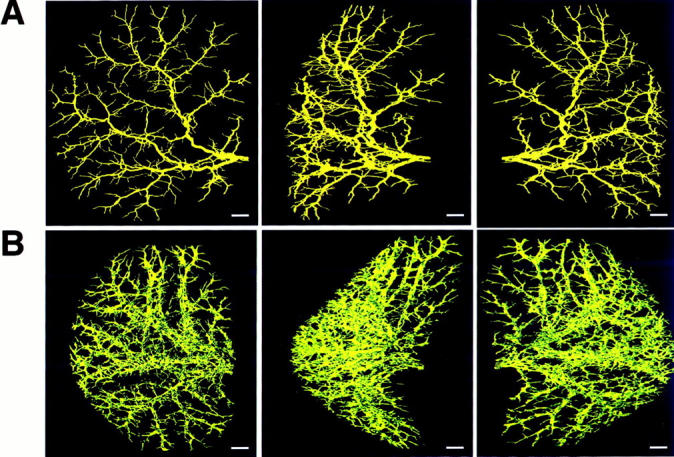
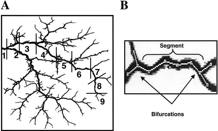
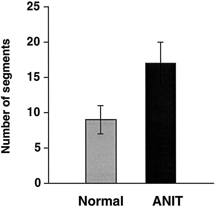
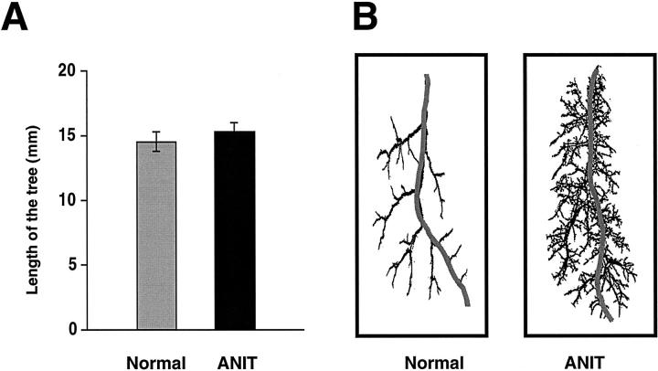
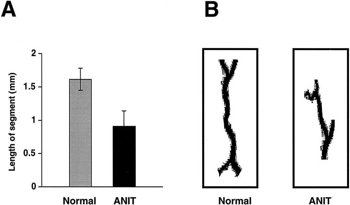
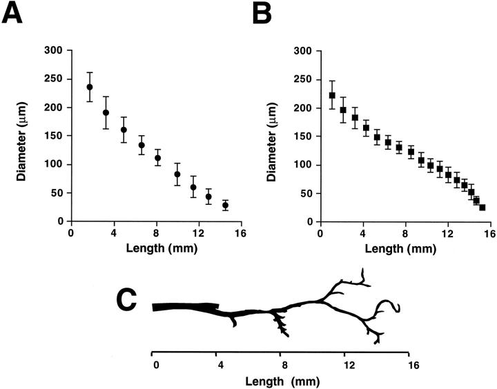
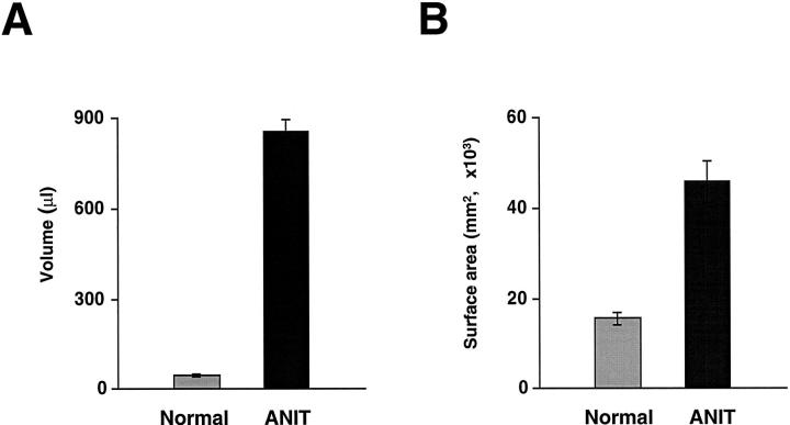
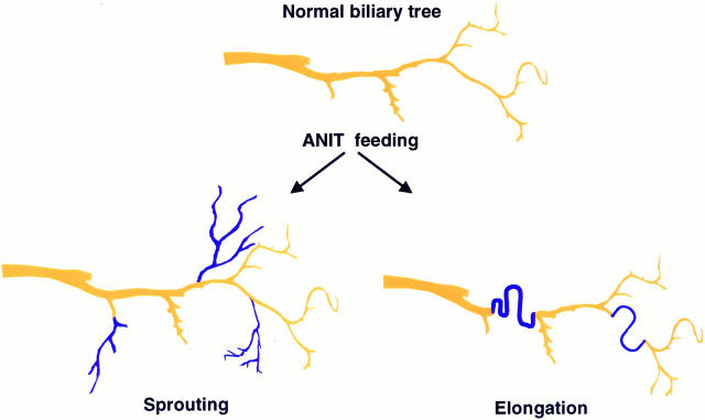
The anatomical details of the biliary tree architecture of normal rats and rats in whom selective proliferation was induced by feeding alpha-naphthylisothiocyanate (ANIT) were reconstructed in three dimension using a microscopic-computed tomography scanner. The intrahepatic biliary tree was filled with a silicone polymer through the common bile duct and each liver lobe embedded in Bioplastic; specimens were then scanned by a microscopic-computed tomography scanner and modified Feldkamp cone beam backprojection algorithm applied to generate three-dimensional images. Quantitative analysis of bile duct geometry was performed using a customized software program. The diameter of the bile duct segments of normal and ANIT-fed rats progressively decreased with increasing length of the biliary tree. Diameter of bile ducts from ANIT-fed rats (range, 21 to 264 microm) was similar to that of normal rats (22 to 279 microm). In contrast, the number of bile duct segments along the major branch reproducibly doubled, the length of the bile duct segments decreased twofold, and the length of the biliary tree remained unchanged after ANIT feeding. Moreover, the total volume of the biliary tree of ANIT-fed rats was significantly greater (855 microl) than in normal rats (47 microl). Compared with normal rats, the total surface area of the biliary tree increased 26 times after ANIT-induced bile duct proliferation. Taken together, these observations quantitate the anatomical remodeling after selective cholangiocyte proliferation and strongly suggest that the proliferative process involves sprouting of new side branches. Our results may be relevant to the mechanisms by which ducts proliferate in response to hepatic injury and to the hypercholeresis that occurs after experimentally induced bile duct proliferation.


Figures

Figure 1.
Figure 1.
The brightest voxel projections of the three-dimensional microCT images of the intrahepatic biliary tree of the left lateral lobe of normal (A) and ANIT-fed (B) rats. Contrast agent for biliary tree visualization was injected into the common bile duct as described in detail in Materials and Methods. Both biliary trees are viewed at 80° increments. Scale bars, 1 mm.
Figure 2.
Figure 2.
Quantitative analysis of the intrahepatic biliary tree. A: An example for quantitative analysis is shown for the biliary tree of normal rat. The major branch of the biliary tree was selected, manually traced, and divided into segments at each branch point. Segments were numbered consecutively from 1 to 9 beginning at the largest bile duct and moving along the major branch to the smallest bile duct. B: Portion of the manually traced major branch. A bifurcation is defined as a point along the biliary tree at which the bile duct divides and a segment as that part of the biliary tree between bifurcations.
Figure 3.
Figure 3.
The number of bile duct segments along the major branch of the intrahepatic biliary tree of normal and ANIT-fed rats. After bile duct proliferation induced by ANIT feeding, the number of bile duct segments was reproducibly doubled (P < 0.001).
Figure 4.
Figure 4.
Length of the intrahepatic biliary tree along the major branch of normal and ANIT-fed rats. A: Length of the biliary tree was calculated as a sum of length of the individual bile duct segments along the major branch. After bile duct proliferation induced by ANIT, the biliary tree length was not different from the length of the biliary tree of normal rats. B: Representative traced major branch of the biliary tree of normal and ANIT-fed rats.
Figure 5.
Figure 5.
Length of the individual bile duct segments from normal and ANIT-fed rats. A: The length of the bile duct segments was measured as a straight-line distance between bifurcations. Bile duct segments from intrahepatic biliary tree of normal rats were two times longer than bile duct segments of ANIT-fed rats (P < 0.001). B: Representative individual bile duct segments from normal and ANIT-fed rats.
Figure 6.
Figure 6.
Diameter of bile duct segments along the major branch of the intrahepatic biliary tree of normal (A) and ANIT-fed (B) rats. Diameters of the individual bile duct segment are shown versus length of the biliary tree along the major branch. Values are mean ± SE (P < 0.05). Note that proliferating bile ducts from ANIT-fed rats have a diameter similar to that of normal bile duct segments. C: Representative of the major branch of the intrahepatic biliary tree. With increasing length of the biliary tree diameter of bile duct segments becomes smaller.
Figure 7.
Figure 7.
Total volumes (A) and total surface areas (B) of the entire biliary tree from normal and ANIT-fed rats. Total volume and total surface area of the biliary tree after bile duct proliferation provoked by ANIT feeding was 18 and 26 times larger, respectively, than if seen in normal rats.
Figure 8.
Figure 8.
Schematic representation of two different types of anatomical remodeling after selective cholangiocyte proliferation induced by ANIT. Under normal conditions, biliary tree regularly branches every 1.5 mm along its main course. After ANIT feeding proliferation of bile duct may occur by sprouting of new side branches or by elongation of the pre-existing bile duct. (Newly formed bile ducts are shown in blue). Proliferation of bile ducts is associated with anatomical alterations in biliary tree architecture. By analysis of these changes, we concluded that biliary tree proliferation results from sprouting of new side branches.

References

    1. LaRusso NF: Morphology, physiology and biochemistry of biliary epithelia. Toxicol Pathol 1996, 24:84-89 - PubMed
    1. Roberts SK, Ludwig J, LaRusso NF: The pathobiology of biliary epithelia. Gastroenterology 1997, 112:269-279 - PubMed
    1. Alpini G, Phillips JO, LaRusso NF: The biology of biliary epithelia. Arias IM Boyer JL Fausto N Jakoby WB Schachter DA Shafritz DA eds. The Liver: Biology and Pathobiology. 1994, :pp 623-653 Ltd., New York, Raven Press
    1. Birnbaum A, Suchy FJ: The intrahepatic cholangiopathies. Semin Liver Dis 1998, 18:263-269 - PubMed
    1. Roberts SK, Kuntz SM, Gores GJ, LaRusso NF: Regulation of bicarbonate-dependent ductular bile secretion assessed by lumenal micropuncture of isolated rodent intrahepatic bile ducts. Proc Natl Acad Sci USA 1993, 90:9080-9084 - PMC - PubMed

Publication types

Substances

LinkOut - more resources