. 2001 Jun;158(6):2127-38.
doi: 10.1016/s0002-9440(10)64684-6.

Myelin/axonal pathology in interleukin-12 induced serial relapses of experimental allergic encephalomyelitis in the Lewis rat

Affiliations

Myelin/axonal pathology in interleukin-12 induced serial relapses of experimental allergic encephalomyelitis in the Lewis rat

Z Ahmed et al. Am J Pathol. 2001 Jun.

Erratum in

  • Am J Pathol 2001 Nov;159(5):1975

Abstract

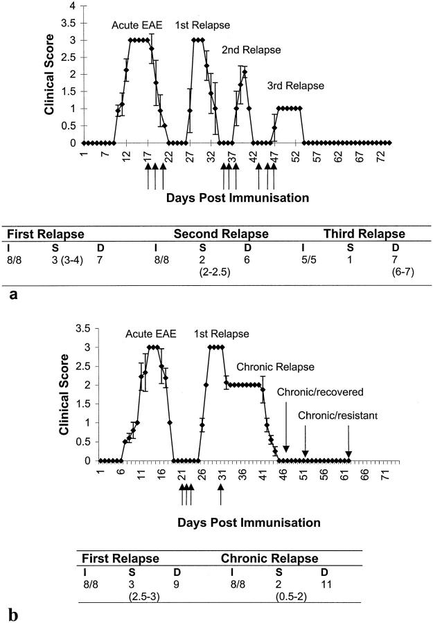
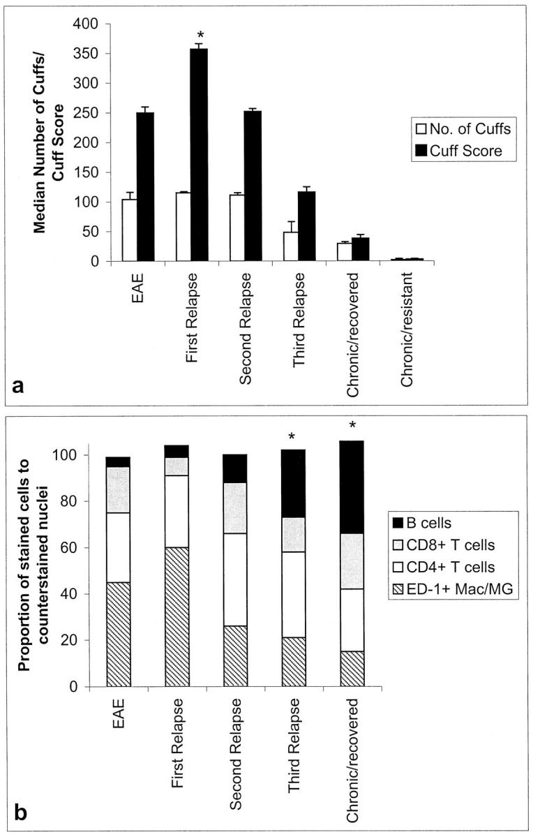
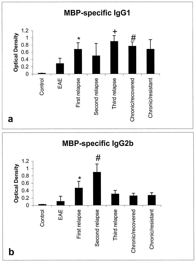
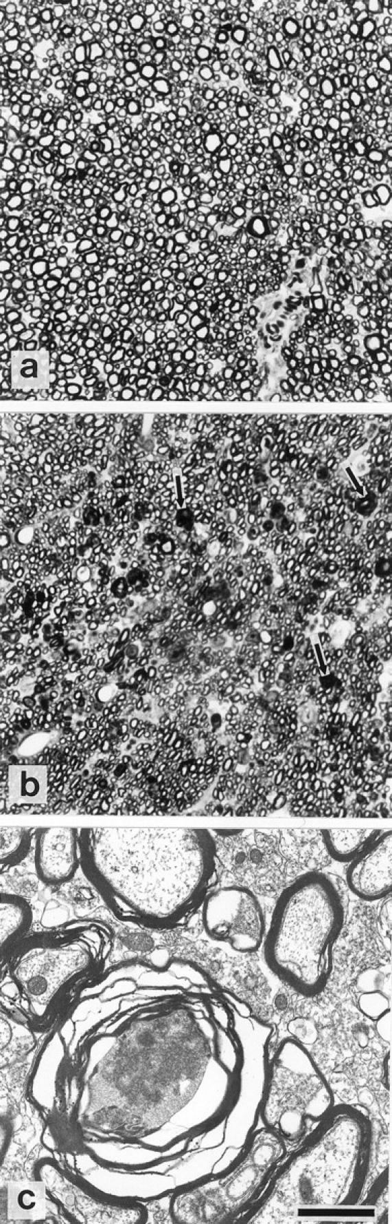
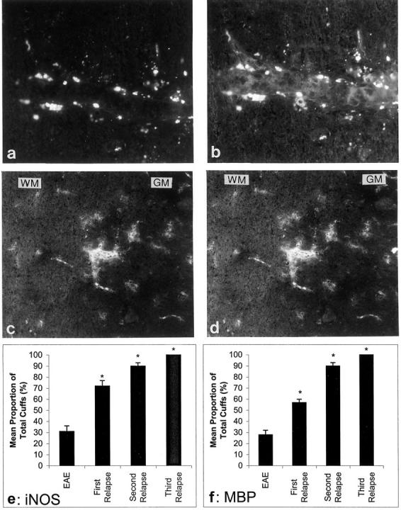
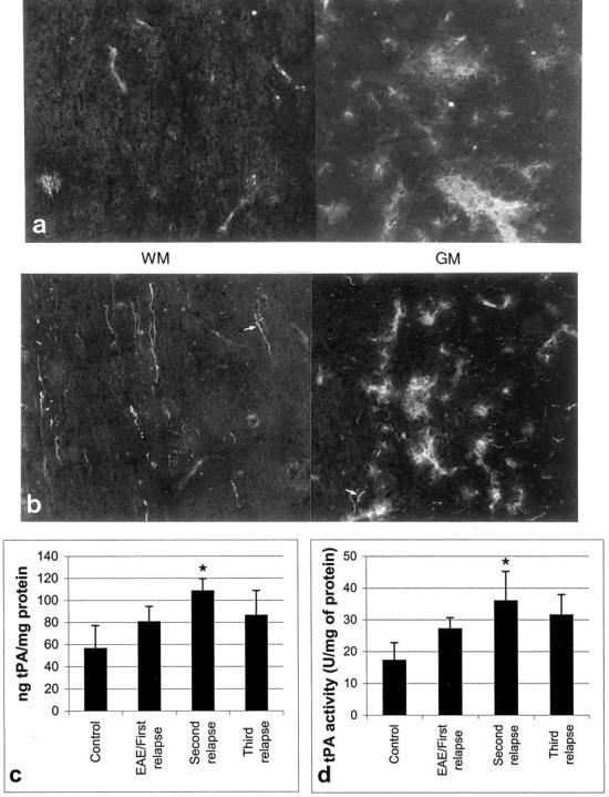
Lewis rats, on recovery from monophasic clinical experimental allergic encephalomyelitis (EAE), can be induced to develop repeated paralytic relapses with a graded reduction in clinical severity following intraperitoneal administration of IL-12. By the time of the third relapse, the number and size of inflammatory cuffs in the spinal cord were reduced with the makeup of the cellular infiltrate shifting to a significantly increased number of B cells. Serum levels of myelin basic protein (MBP)-specific IgG1 and IgG2b were found to rise over time while MBP and MBP peptide-positive macrophages and microglia became evident in perivascular cuffs and in spinal cord parenchyma, indicative of myelin phagocytosis. Axonal death was observed in semithin and EM sections of spinal cord in third relapse animals in association with iNOS and tPA immunostaining throughout gray and white matter. These neurotoxic or excitotoxic agents may contribute to axonal damage directly or indirectly by activated microglia and macrophages, leading to limited damage of the axonal-myelin unit.


Figures

Figure 1.
Figure 1.
IL-12 induces serial paralytic relapses in EAE with diminishing severity. a: IL-12 (3 μg/ml, 0.2 ml i.p.) was injected (arrows) on days 17, 19, 21, days 35, 37, and 39 for second relapse, and days 45, 47, and 49 for third relapse (indicated by arrows). Points represent the mean ± SD of clinical scores. The table represents the: I, incidence; S, severity; and D, duration of disease for each relapse. Values represent median and range. b: Chronic relapse experiment where IL-12 was injected as for the first relapse (days 21, 23, and 25) followed by allowing the animals to recover to clinical score 2 before being injected with IL-12 at a dose of 1.5 μg/ml, 0.2 ml, i.p. every other day (days 35, 37, and 39 followed by every other day until day 63).
Figure 2.
Figure 2.
CNS histopathology in IL-12-induced relapses. Graphs show cuff numbers and score (a) and the cellular distribution of the inflammatory cuffs (b). *P < 0.001 versus EAE. Mac, macrophages; MG, microglia.
Figure 3.
Figure 3.
B cell localization in inflammatory cuffs in cervical regions of the spinal cord in acute EAE (a), first relapse (b), second relapse (c), and third relapse animals (d). Original magnification, ×1000.
Figure 4.
Figure 4.
Biologically active TGF-β levels increase during EAE and relapses. ***P < 0.01.
Figure 5.
Figure 5.
IgG1 and IgG2b isotype responses showed a general increase in levels of MBP-specific IgG1 (*P < 0.001 EAE versus first relapse, +P < 0.001 first relapse versus third relapse, #P < 0.001 first relapse versus chronic/recovered) (a) and MBP-specific IgG2b (*P < 0.05 EAE versus first relapse, #P < 0.05 first relapse versus second relapse) (b) after IL-12-induced relapses.
Figure 6.
Figure 6.
Axonal death in the dorsal funiculi region of the spinal cord during the third relapse (b: representative cervical region shown) indicated by arrows. Note the smaller diameter of axons in the damaged region (b) compared to the identical region in untreated control animals (a). c: EM showing degenerating axon surrounded by disrupted myelin. Original magnification in a and b, ×1000; bar in c = 1 μm.
Figure 7.
Figure 7.
Representative MBP and iNOS immunostaining localized in macrophages in the spinal cord of Lewis rats. Double staining on serial sections with ED-1 (a) and MBP (b). c: iNOS immunostaining was closely linked to the pattern of MBP (serial sections) (d). *P < 0.001 versus EAE. Original magnifications, ×500. Graphs to show the proportion of inflammatory cuffs which were positive for iNOS (e) and MBP (f).
Figure 8.
Figure 8.
A composite photomicrograph of neighboring frames to show tPA expression in macrophages of inflammatory cuffs in spinal cord WM and GM in first relapse (a) and in axons of third relapse (b) animals. Original magnifications, ×500. Total tPA protein levels (c) as well as tPA activity (d) increased during the second relapse (*P < 0.001 versus control).

References

    1. Lassman H, Zimprich F, Rossier K, Vass K: Inflammation in the central nervous system: basic mechanisms and immunological concepts. Rev Neurol 1991, 147:763-781 - PubMed
    1. Ben-Nun A, Cohen I: Experimental autoimmune encephalomyelitis (EAE) mediated by T cell lines: process of selection of lines and characterization of the cells. J Immunol 1982, 129:303-308 - PubMed
    1. Huitinga I, Van Roojien N, de Groot CJA, Uitdehaag BMJ, Dijkstra CD: Suppression of experimental allergic encephalomyelitis in Lewis rats after elimination of macrophages. J Exp Med 1990, 172:1025-1033 - PMC - PubMed
    1. Powell MB, Mitchell D, Lederman J, Buchmeier J, Zamvil SS, Graham M, Ruddle NH, Steinman L: Lymphotoxin production by myelin basic protein specific T cell clones correlate with encephalitogenicity. Int Immunol 1990, 2:539-544 - PubMed
    1. Sedgwick JD, McPhee JHM, Puklawec M: Isolation of encephalitogenic CD4+ T cell clones in the rat: cloning methodology and IFN-γ secretion. J Immunol Methods 1989, 143:3492-3497 - PubMed

Publication types

MeSH terms

LinkOut - more resources