Dexamethasone inhibits TNF-alpha-induced apoptosis and IAP protein downregulation in MCF-7 cells
- PMID: 11399663
- PMCID: PMC1572806
- DOI: 10.1038/sj.bjp.0704093
Dexamethasone inhibits TNF-alpha-induced apoptosis and IAP protein downregulation in MCF-7 cells
Abstract
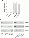
Exposure of human mammary carcinoma cell line MCF-7 to TNF-alpha leads to apoptotic cell death within 24 h. In search for apoptosis-preventing signals, we identified glucocorticoids as potent death-preventing compounds. Ten nM dexamethasone provided a significant protective effect whereas 100 nM dexamethasone roughly blocked 80 - 90% of TNF-alpha-induced apoptosis. Surprisingly, dexamethasone exerted a protective effect even when supplied several hours after TNF-alpha. This points to a powerful inhibition of even advanced apoptotic processes by dexamethasone. To further pinpoint the anti-apoptotic glucocorticoid action, we investigated the expression levels of several members of the inhibitors of apoptosis (IAPs) family of proteins in response to TNF-alpha and dexamethasone. IAP proteins directly block caspase protease activities including caspase-3, caspase-7, and caspase-9. Exposure of MCF-7 cells to TNF caused an extensive downregulation of cIAP1, cIAP2, and XIAP protein levels. The decline of the IAP protein levels temporally paralleled the appearance of apoptotic DNA fragments which started 12 - 14 h following TNF-alpha addition and maximal effects were seen within 24 h. Coincubation of cells with TNF-alpha and dexamethasone potently blocked cIAP1, cIAP2, and XIAP downregulation. TNF-alpha-mediated IAP protein downregulation was not affected by proteasome inhibitors like lactacystin, ALLN or ALLM, whereas it was blocked by the broad-spectrum caspase inhibitor Z-VAD-fmk which also prevented TNF-alpha-induced apoptotic cell death. These data suggest that inhibition of IAP downregulation mediated by a caspase proteolytic activity constitutes the anti-apoptotic action of glucocorticoids in MCF-7 carcinoma cells.
Figures


Similar articles
-
Suppression of apoptosis by glucocorticoids in glomerular endothelial cells: effects on proapoptotic pathways.Br J Pharmacol. 2000 Apr;129(8):1673-83. doi: 10.1038/sj.bjp.0703255. Br J Pharmacol. 2000. PMID: 10780973 Free PMC article.
-
Dexamethasone protection from TNF-alpha-induced cell death in MCF-7 cells requires NF-kappaB and is independent from AKT.BMC Cell Biol. 2006 Feb 21;7:9. doi: 10.1186/1471-2121-7-9. BMC Cell Biol. 2006. PMID: 16504042 Free PMC article.
-
c-IAP1 blocks TNFalpha-mediated cytotoxicity upstream of caspase-dependent and -independent mitochondrial events in human leukemic cells.Biochem Biophys Res Commun. 2001 Sep 14;287(1):181-9. doi: 10.1006/bbrc.2001.5582. Biochem Biophys Res Commun. 2001. PMID: 11549272
-
Regulation of TRAIL-induced apoptosis by ectopic expression of antiapoptotic factors.Vitam Horm. 2004;67:453-83. doi: 10.1016/S0083-6729(04)67023-3. Vitam Horm. 2004. PMID: 15110190 Review.
-
Inhibitors of apoptotic proteins: new targets for anticancer therapy.Chem Biol Drug Des. 2013 Sep;82(3):243-51. doi: 10.1111/cbdd.12176. Chem Biol Drug Des. 2013. PMID: 23790005 Review.
Cited by
-
Influenza B Virus (IBV) Immune-Mediated Disease in C57BL/6 Mice.Vaccines (Basel). 2022 Sep 1;10(9):1440. doi: 10.3390/vaccines10091440. Vaccines (Basel). 2022. PMID: 36146518 Free PMC article.
-
Selective glucocorticoid receptor-activating adjuvant therapy in cancer treatments.Oncoscience. 2016 Jul 27;3(7-8):188-202. doi: 10.18632/oncoscience.315. eCollection 2016. Oncoscience. 2016. PMID: 27713909 Free PMC article. Review.
-
Hypothalamic-Pituitary-Adrenal Hormones Impair Pig Fertilization and Preimplantation Embryo Development via Inducing Oviductal Epithelial Apoptosis: An In Vitro Study.Cells. 2022 Dec 2;11(23):3891. doi: 10.3390/cells11233891. Cells. 2022. PMID: 36497149 Free PMC article.
-
The molecular mechanisms of glucocorticoids-mediated neutrophil survival.Curr Drug Targets. 2011 Apr;12(4):556-62. doi: 10.2174/138945011794751555. Curr Drug Targets. 2011. PMID: 21504070 Free PMC article. Review.
-
Inhibition of caspase mediated apoptosis restores muscle function after crush injury in rat skeletal muscle.Apoptosis. 2012 Mar;17(3):269-77. doi: 10.1007/s10495-011-0674-1. Apoptosis. 2012. PMID: 22089165 Free PMC article.
References
-
- BALLERMANN B.J. Regulation of bovine glomerular endothelial cell growth in vitro. Am. J. Physiol. 1989;256:C182–189. - PubMed
-
- BAKER S.J., REDDY P. Transducers of life and death: TNF receptor superfamily and associated proteins. Oncogene. 1996;12:1–9. - PubMed
-
- BEG A.A., BALTIMORE D. An essential role for NF-κB in preventing TNF-α-induced cell death. Science. 1996;274:782–784. - PubMed
-
- BRADFORD M.M. A rapid and sensitive method for the quantification of protein utilizing the principle of protein-dye binding. Anal. Biochem. 1976;72:248–254. - PubMed
Publication types
MeSH terms
Substances
LinkOut - more resources
Full Text Sources
Other Literature Sources
Research Materials

