

Calprotectin expression in vitro by oral epithelial cells confers resistance to infection by Porphyromonas gingivalis
- PMID: 11401960
- PMCID: PMC98457
- DOI: 10.1128/IAI.69.7.4242-4247.2001
Calprotectin expression in vitro by oral epithelial cells confers resistance to infection by Porphyromonas gingivalis
Abstract
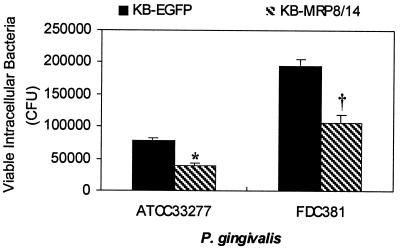
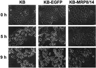
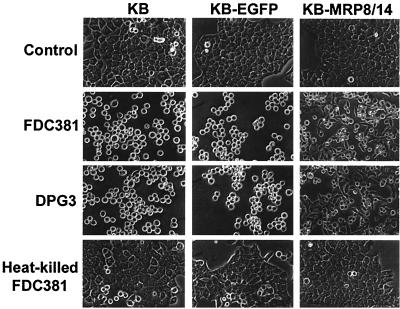
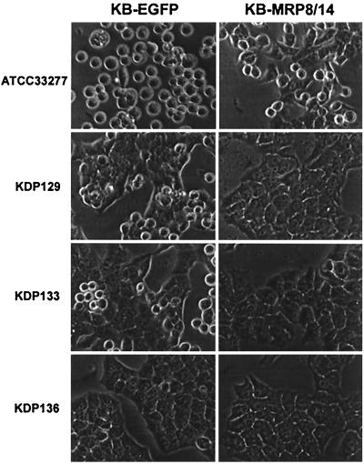
Calprotectin, an S100 calcium-binding protein with broad-spectrum antimicrobial activity in vitro, is expressed in neutrophils, monocytes, and gingival keratinocytes. In periodontitis, calprotectin appears upregulated and is detected at higher levels in gingival crevicular fluid and tissue specimens. How calprotectin contributes to the pathogenesis of periodontal diseases is unknown. To isolate the effects of calprotectin, a calprotectin-negative oral epithelial cell line was transfected with calprotectin genes to enable expression. Porphyromonas gingivalis was permitted to bind and invade transfected cells expressing calprotectin and sham transfectants. Rates of invasion into both cell lines were compared using the antibiotic protection assay. Transfected cells expressing calprotectin showed 40 to 50% fewer internalized P. gingivalis than sham transfectants. Similarly, binding to calprotectin expressing cells was reduced approximately twofold at all time points (15, 30, 45, and 60 min) as estimated by immunofluorescence analysis. Independent of invasion, however, prolonged exposure to P. gingivalis induced epithelial cell rounding and detachment from the substratum. These morphological changes were delayed, however, in cells expressing calprotectin. Using P. gingivalis protease-deficient mutants, we found that Arg-gingipain and Lys-gingipain contributed to epithelial cell rounding and detachment. In conclusion, expression of calprotectin appears to protect epithelial cells in culture against binding and invasion by P. gingivalis. In addition, cells expressing calprotectin are more resistant to detachment mediated by Arg-gingipain and Lys-gingipain. In periodontal disease, calprotectin may augment both the barrier protection and innate immune functions of the gingival epithelium to promote resistance to P. gingivalis infection.
Figures

References
-
- Belton C M, Izutsu K T, Goodwin P C, Park Y, Lamont R J. Fluorescence image analysis of the association between Porphyromonas gingivalis and gingival epithelial cells. Cell Microbiol. 1999;1:215–233. - PubMed
-
- Bhardwaj R S, Zotz C, Zwadlo-Klarwasser G, Roth J, Goebeler M, Mahnke K, Falk M, Meinardus-Hager G, Sorg C. The calcium-binding proteins MRP8 and MRP14 form a membrane-associated heterodimer in a subset of monocytes/macrophages present in acute but absent in chronic inflammatory lesions. Eur J Immunol. 1992;22:1891–1897. - PubMed
-
- Brandtzaeg P, Dale I, Fagerhol M K. Distribution of a formalin-resistant myelomonocytic antigen (L1) in human tissues. IL. Normal and aberrant occurrence in various epithelia. Am J Clin Pathol. 1987;87:700–707. - PubMed
-
- Brandtzaeg P, Gabrielsen T O, Dale I, Muller F, Steinbakk M, Fagerhol M K. The leucocyte protein L1 (calprotectin): a putative nonspecific defence factor at epithelial surfaces. Adv Exp Med Biol. 1995;371A:201–206. - PubMed
Publication types
MeSH terms
Substances
Grants and funding
LinkOut - more resources
Full Text Sources
Other Literature Sources
