. 2001 Jul;69(7):4610-7.
doi: 10.1128/IAI.69.7.4610-4617.2001.

Salmonella-induced cell death is not required for enteritis in calves

Affiliations

Salmonella-induced cell death is not required for enteritis in calves

R L Santos et al. Infect Immun. 2001 Jul.

Abstract

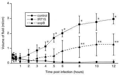
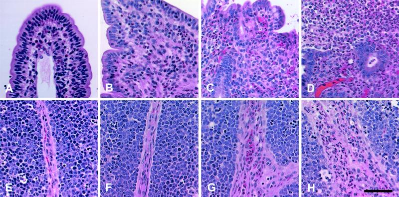
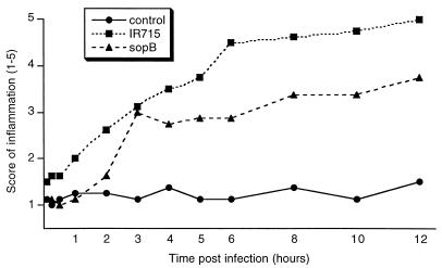
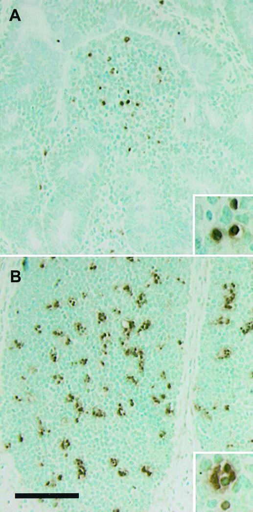
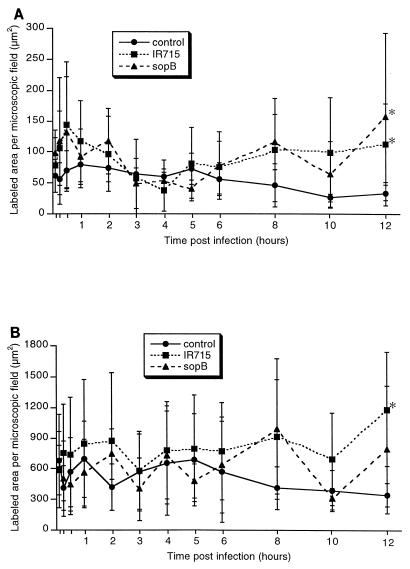
Salmonella enterica serovar Typhimurium causes cell death in bovine monocyte-derived and murine macrophages in vitro by a sipB-dependent mechanism. During this process, SipB binds and activates caspase-1, which in turn activates the proinflammatory cytokine interleukin-1beta through cleavage. We used bovine ileal ligated loops to address the role of serovar Typhimurium-induced cell death in induction of fluid accumulation and inflammation in this diarrhea model. Twelve perinatal calves had 6- to 9-cm loops prepared in the terminal ileum. They were divided into three groups: one group received an intralumen injection of Luria-Bertani broth as a control in 12 loops. The other two groups (four calves each) were inoculated with 0.75 x 10(9) CFU of either wild-type serovar Typhimurium (strain IR715) or a sopB mutant per loop in 12 loops. Hematoxylin and eosin-stained sections were scored for inflammation, and terminal deoxynucleotidyltransferase-mediated dUTP-biotin nick end labeling (TUNEL)-positive cells were detected in situ. Fluid accumulation began at 3 h postinfection (PI). Inflammation was detected in all infected loops at 1 h PI. The area of TUNEL-labeled cells in the wild-type infected loops was significantly higher than that of the controls at 12 h PI, when a severe inflammatory response and tissue damage had already developed. The sopB mutant induced the same amount of TUNEL-positive cells as the wild type, but it was attenuated for induction of fluid secretion and inflammation. Our results indicate that serovar Typhimurium-induced cell death is not required to trigger an early inflammatory response and fluid accumulation in the ileum.


Figures

FIG. 1
FIG. 1
Time course of fluid accumulation into the ileal lumen during serovar Typhimurium infection from 5 min through 12 h postinfection. Each data point represents the average (± standard deviation) of four independent experiments. ∗, Values corresponding to wild-type infected loops (IR715) are significantly higher than those for the uninfected controls and loops infected with sopB mutant from 3 to 12 h postinfection (P < 0.05). ∗∗, Values corresponding to sopB-infected loops are significantly higher than those for uninfected controls and significantly lower than wild-type infected loops (IR715) from 8 to 12 h postinfection (P < 0.05).
FIG. 2
FIG. 2
Micrographs of bovine Peyer's patches infected with wild-type serovar Typhimurium. (A to D) Sections of the mucosa. (A) Uninfected control, no significant histological changes; (B) at 1 h postinfection, mild focal infiltration of neutrophils; (C) mucosa at 6 h postinfection, diffuse infiltration of neutrophils and marked blunting of the villi; (D) at 12 h postinfection, severe diffuse infiltration of neutrophils with loss of superficial epithelium and extensive exudation into the lumen. (E to H) Sections of lymphoid nodules. (E) uninfected control, no significant histological changes; (F) at 1 h postinfection, with no significant histological changes; (G) at 6 h post infection, perivascular infiltration of neutrophils in the interstitial connective tissue; (H) at 12 h postinfection, severe diffuse infiltration of neutrophils in the interstitial connective tissue. Stain is hematoxylin and eosin. Bar = 50 μm.
FIG. 3
FIG. 3
Inflammatory changes in the Peyer's patches after serovar Typhimurium infection. Magnitude of inflammation was scored ( to 5) according to the criteria described in Materials and Methods. Each data point represents the average of four independent experiments.
FIG. 4
FIG. 4
Localization of TUNEL-positive cells. In sections of Peyer's patches at 2 h after infection with wild-type serovar Typhimurium, TUNEL-stained cells were more concentrated in the domed villi (A) and lymphoid nodules (B). Methyl green counterstaining was used. Bar = 100 μm.
FIG. 5
FIG. 5
Area of in situ TUNEL labeling per microscopic field in the Peyer's patches during serovar Typhimurium infection. The area of labeling per 36,500-μm2 microscopic field was measured using the NIH Image software as described in Materials and Methods. Each data point represents the average ± standard deviation of four independent experiments. (A) Mucosa; ∗ indicates differences between control and loops infected with either the wild type (IR715) or sopB mutant at 12 h postinfection are statistically significant (P < 0.05). (B) Lymphoid nodules; ∗ indicates the difference between control and wild-type (IR715) infected loops at 12 h postinfection is statistically significant (P < 0.05).
FIG. 6
FIG. 6
Flow cytometric analysis of bovine macrophages infected with wild-type serovar Typhimurium or of sopB mutant grown to the logarithmic phase. Macrophages were infected in Teflon flasks with multiplicity of infection of 50:1, harvested 1 h after infection, processed for TUNEL staining (see Materials and Methods), and analyzed by flow cytometry. In each dot plot, the x axis corresponds to propidium iodide staining and the y axis corresponds to Br-dUTP incorporation. TUNEL-positive cells are within the area indicated by the quadrilateral, and the percentage of these cells is indicated at the left top corner of each panel. These data are from a representative experiment showing uninfected macrophages with a low background of TUNEL-positive cells (A) or macrophages infected with the wild type (B) or sopB mutant (C) containing a high percentage of apoptotic cells.
FIG. 7
FIG. 7
Invasion of bovine Peyer's patches by the wild type (black bars) or a sopB mutant (gray bars) of serovar Typhimurium. Tissue samples from Peyer's patches were weighed, homogenized in PBS, serially diluted, and plated onto LB agar plates containing nalidixic acid (50 μg/ml) for counting CFU. Each bar represents the mean and standard deviation of a time point from four independent experiments.

References

    1. Ahmer B M, van Reeuwijk J, Watson P R, Wallis T S, Heffron F. Salmonella SirA is a global regulator of genes mediating enteropathogenesis. Mol Microbiol. 1999;31:971–982. - PubMed
    1. Baran J, Guzik K, Hryniewicz W, Ernst M, Flad H D, Pryjma J. Apoptosis of monocytes and prolonged survival of granulocytes as a result of phagocytosis of bacteria. Infect Immun. 1996;64:4242–4248. - PMC - PubMed
    1. Brennan M A, Cookson B T. Salmonella induces macrophage death by caspase-1-dependent necrosis. Mol Microbiol. 2000;38:31–40. - PubMed
    1. Chen L M, Kaniga K, Galán J E. Salmonella spp. are cytotoxic for cultured macrophages. Mol Microbiol. 1996;21:1101–1115. - PubMed
    1. Conover W J. Practical nonparametric statistics. New York, N.Y: Wiley; 1980.

Publication types