. 2001 Jun 11;153(6):1199-208.
doi: 10.1083/jcb.153.6.1199.

HCP-4, a CENP-C-like protein in Caenorhabditis elegans, is required for resolution of sister centromeres

Affiliations

HCP-4, a CENP-C-like protein in Caenorhabditis elegans, is required for resolution of sister centromeres

L L Moore et al. J Cell Biol. .

Abstract

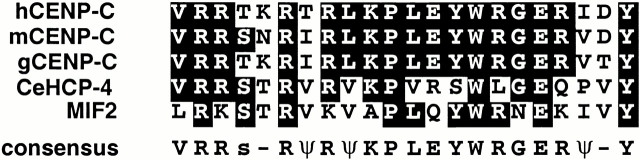
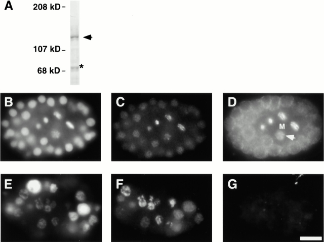
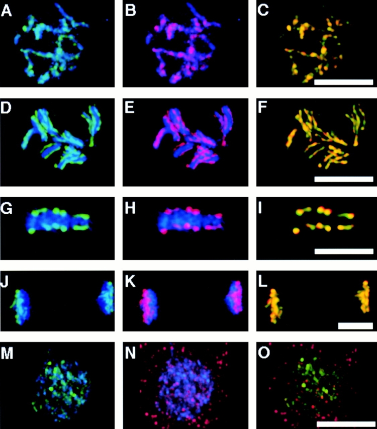
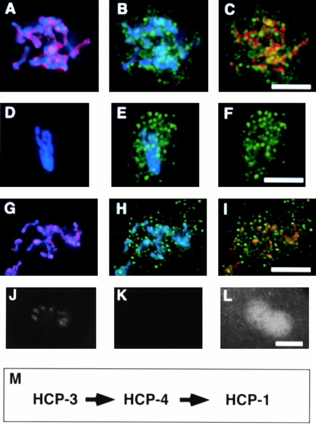
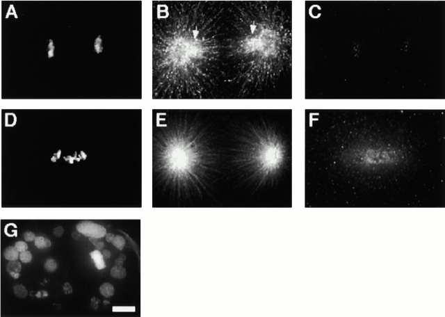
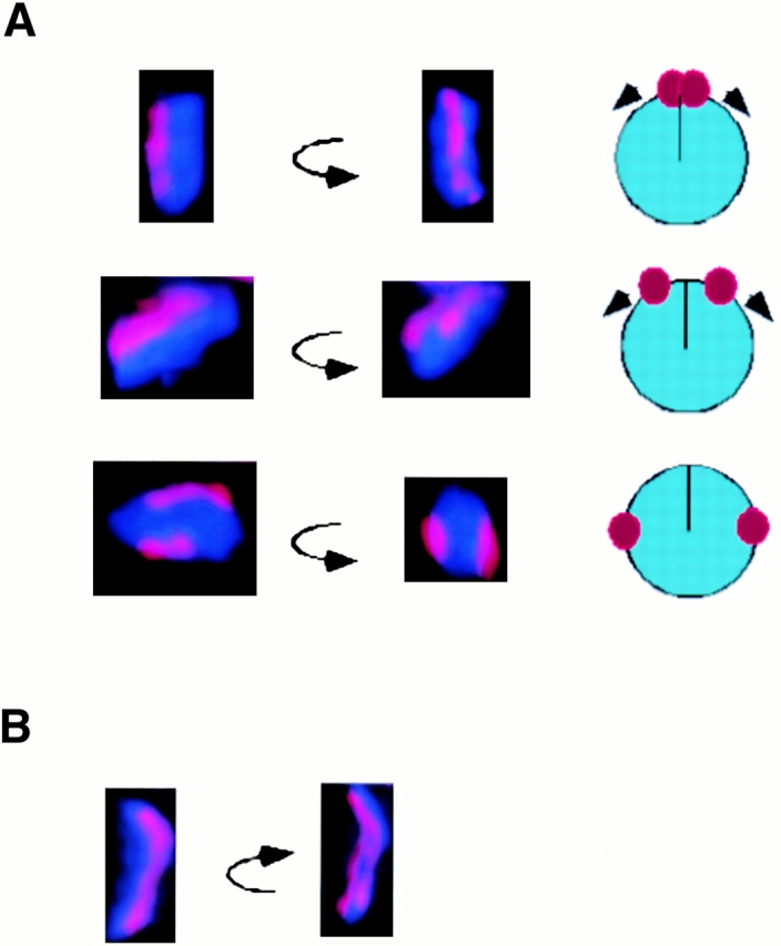
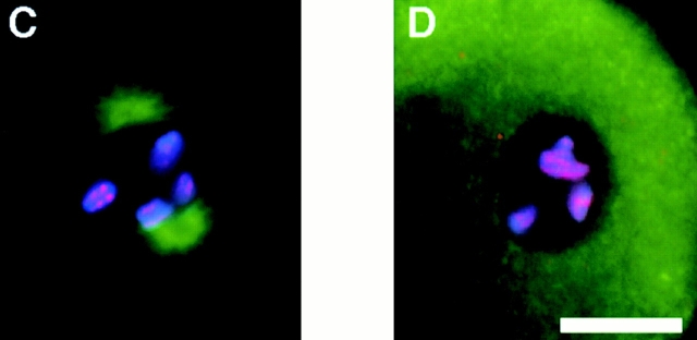
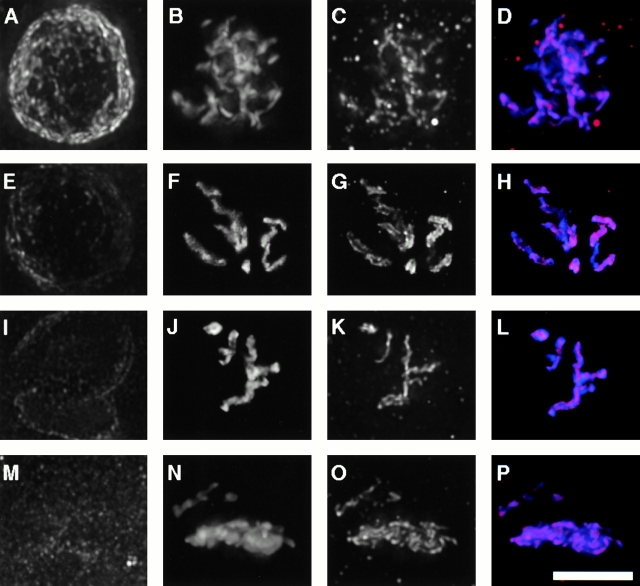
The centromere plays a critical role in the segregation of chromosomes during mitosis. In mammals, sister centromeres are resolved from one another in the G2 phase of the cell cycle. During prophase, chromosomes condense with sister centromeres oriented in a back to back configuration enabling only one chromatid to be captured by each half spindle. To study this process, we identified a centromere protein (CENP)-C-like protein, holocentric protein (HCP)-4, in Caenorhabditis elegans based on sequence identity, loss of function phenotype, and centromeric localization. HCP-4 is found in the cytoplasm during interphase, but is nuclear localized in mitosis, where it localizes specifically to the centromere. The localization of HCP-4 to the centromere is dependent on the centromeric histone HCP-3; in addition, HCP-3 and HCP-4 are both required for localization of a CENP-F-like protein, HCP-1, indicating an ordered assembly pathway. Loss of HCP-4 expression by RNA-mediated interference resulted in a failure to generate resolution of sister centromeres on chromosomes, suggesting that HCP-4 is required for sister centromere resolution. These chromosomes also failed to form a functional kinetochore. Thus, the CENP-C-like protein HCP-4 is essential for both resolution sister centromeres and attachment to the mitotic spindle.


Figures

Figure 1
Figure 1
Sequence and alignment of HCP-4 with CENP-C proteins. BLOCKMAKER and the Motif Alignment and Search Tool programs were used with human CENP-C (EMBL/GenBank/DDBJ accession no. M95724), mouse CENP-C (EMBL/GenBank/DDBJ accession no. U03113), chicken CENP-C (EMBL/GenBank/DDBJ accession no. AB004649), and MIF2 (EMBL/GenBank/DDBJ accession no. Z28089) to identify a region of similarity in HCP-4 (Bailey and Gribskov 1998; Henikoff and Henikoff 1991). Alignment was performed using the ClustalW multiple alignment program (Thompson et al. 1994) and displayed using Boxshade (http://www.ch.embnet.org/software/BOX_form.html). Identities are shaded black.
Figure 2
Figure 2
HCP-4 localization in C. elegans embryos. (A) Western blot of wild-type embryo extract (60 μg total protein) probed with α–HCP-4 antibody (diluted 1:500) shows that a single band of ∼136 kD is present. All other bands, including the band at 70 kD (*), were independent of the primary antibody. Wild-type embryos or hcp-4(RNAi) embryos were stained with DAPI (B and E), α–HCP-3 antibody (C and F), and α–HCP-4 antibody (D and G). Approximately equal staged embryos containing both mitotic (M) and interphase cells are shown. Exposure times were normalized relative to the HCP-3 staining. Arrow indicates prophase nucleus with nuclear staining. Bar, 5 μm.
Figure 3
Figure 3
Centromeric association of HCP-4 during mitosis. Two-cell C. elegans embryos at different stages of the mitotic cell cycle were fixed and stained with α-HCP3 antibody (A, D, G, J, and M, green), α-HCP4 (B, E, H, K, and N, red), and DAPI (blue). Colocalization of HCP-3 and HCP-4 was visualized in the merged images as yellow (C, F, I, L, and O). All images, except metaphase, are a projection of the Z-stack of the entire nucleus. (A–C) Early prophase; (D–F) late prophase; (G–I) metaphase; (J–L) anaphase; (M–O) telophase. Bars, 5 μm.
Figure 5
Figure 5
The centromere is assembled during prophase. Nuclei from wild-type (A–C), hcp-3(RNAi) (D–F), and hcp-4 (RNAi) (G–I) embryos were fixed and stained with DAPI (blue), α–HCP-3 pAb (red), and α–HCP-1 (green). A P0 hcp-3(RNAi) embryo, postpronuclei fusion, stained with DAPI (J), α–HCP-3 pAb (K), and α–HCP-4 pAb (L). (M) Diagram summarizing the order of assembly for the centromere–kinetochore complex. Bar, 5 μm.
Figure 4
Figure 4
HCP-4 is necessary for kinetochore function. One-cell wild-type (A–C) or hcp-4(RNAi) (D–F) embryos in anaphase were stained with an anti–β-tubulin antibody (B and E), and an α-HCP-1 antibody (C and F). DNA (A and D) was visualized with DAPI. The arrows in B show that the most intensely stained region of the spindle (excluding the centrosomes) is the kinetochore fiber. The kinetochore fiber not observed in hcp-4(RNAi) embryos is shown in E. (G) Late stage hcp-4(RNAi) embryo stained with DAPI, showing a nonuniform distribution of DNA. Bar, 5 μm.
Figure 6
Figure 6
Sister centromere resolution. Individual chromosomes from prophase nuclei were stained with DAPI (blue), α–HCP-1 antibody (not shown), and α–HCP-3 antibody (red). Nuclei containing little or no HCP-1 staining were optically sectioned and a three-dimensional projection generated using the volume viewer function. The resulting images were rotated around the Y-axis. (A) Chromosomes showing different degrees of sister centromere separation. Cartoon of the Z-axis of each chromosome depicting the sister centromeres migration from a juxtaposed position to a maximally resolved orientation is shown adjacent to each image. (B) A single chromosome showing an intermediate “splitting” of sister centromeres. (C and D) Sister centromere resolution is a microtubule-independent process. (C) P1 blastomere from a wild-type embryo stained with DAPI (blue), antitubulin antibody (green), and α–HCP-3 antibody (red). (D) P1 blastomere, from an embryo incubated in the presence of 30 μg/ml nocodazole at room temperature for 30 min, stained as in C. The division time for a two-cell embryo at room temperature is ∼20–30 min, so this embryo had probably just finished cell division when treated with nocodazole. Furthermore, the centrosomes are duplicated (not shown) but have not migrated, indicating that loss of spindle microtubules occurred before centrosome migration, an event that occurs concurrent or before sister centromere resolution (C). Bar, 5 μM.
Figure 6
Figure 6
Sister centromere resolution. Individual chromosomes from prophase nuclei were stained with DAPI (blue), α–HCP-1 antibody (not shown), and α–HCP-3 antibody (red). Nuclei containing little or no HCP-1 staining were optically sectioned and a three-dimensional projection generated using the volume viewer function. The resulting images were rotated around the Y-axis. (A) Chromosomes showing different degrees of sister centromere separation. Cartoon of the Z-axis of each chromosome depicting the sister centromeres migration from a juxtaposed position to a maximally resolved orientation is shown adjacent to each image. (B) A single chromosome showing an intermediate “splitting” of sister centromeres. (C and D) Sister centromere resolution is a microtubule-independent process. (C) P1 blastomere from a wild-type embryo stained with DAPI (blue), antitubulin antibody (green), and α–HCP-3 antibody (red). (D) P1 blastomere, from an embryo incubated in the presence of 30 μg/ml nocodazole at room temperature for 30 min, stained as in C. The division time for a two-cell embryo at room temperature is ∼20–30 min, so this embryo had probably just finished cell division when treated with nocodazole. Furthermore, the centrosomes are duplicated (not shown) but have not migrated, indicating that loss of spindle microtubules occurred before centrosome migration, an event that occurs concurrent or before sister centromere resolution (C). Bar, 5 μM.
Figure 7
Figure 7
HCP-4 is required for sister centromere resolution. Nuclei from wild-type and hcp-4(RNAi) embryos stained with mAb 414 (A, E, I, and M), α–HCP-3 antibody (C, G, K, and O) and DAPI (B, F, J, and N). Merged DAPI (blue) and HCP-3 (red) are shown in (D, H, L, and P). (A–D) Wild-type prophase; (E–H) wild-type prometaphase; (I–L) hcp-4(RNAi) prometa-/metaphase; (M–P) hcp-4(RNAi) anaphase. The cell cycle position was inferred from mAb 414 staining (Lee et al. 2000). Bar, 5 μM.

Comment in

References

    1. Bailey T.L., Gribskov M. Combining evidence using p-valuesapplication to sequence homology searches. Bioinformatics. 1998;14:48–54. - PubMed
    1. Brenner S., Pepper D., Berns M.W., Tan E., Brinkley B.R. Kinetochore structure, duplication, and distribution in mammalian cellsanalysis by human autoantibodies from scleroderma patients. J. Cell Biol. 1981;91:95–102. - PMC - PubMed
    1. Brinkley B.R., Stubblefield E. Ultrastructure and interaction of the kinetchore and centriole in mitosis and meiosis. In: Prescott D.M., Goldstein L., McConkey E., editors. Advances in Cell Biology. Vol. 1. Appleton-Century-Crofts; New York: 1970. pp. 119–185.
    1. Buchwitz B.J., Ahmad K., Moore L.L., Roth M.B., Henikoff S. A histone-H3-like protein in C. elegans . Nature. 1999;401:547–548. - PubMed
    1. Carrington W.A., Lynch R.M., Moore E.D., Isenberg G., Fogarty K.E., Fay F.S. Superresolution three-dimensional images of fluorescence in cells with minimal light exposure. Science. 1995;268:1483–1487. - PubMed

Publication types

MeSH terms

Associated data