. 2001 Jun 11;153(6):1251-64.
doi: 10.1083/jcb.153.6.1251.

A novel integrin-linked kinase-binding protein, affixin, is involved in the early stage of cell-substrate interaction

Affiliations

A novel integrin-linked kinase-binding protein, affixin, is involved in the early stage of cell-substrate interaction

S Yamaji et al. J Cell Biol. .

Abstract

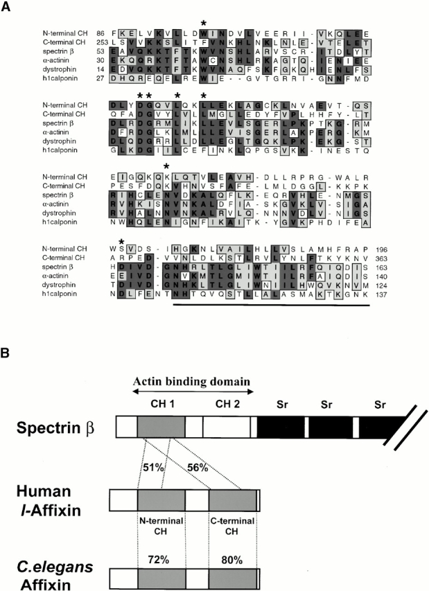
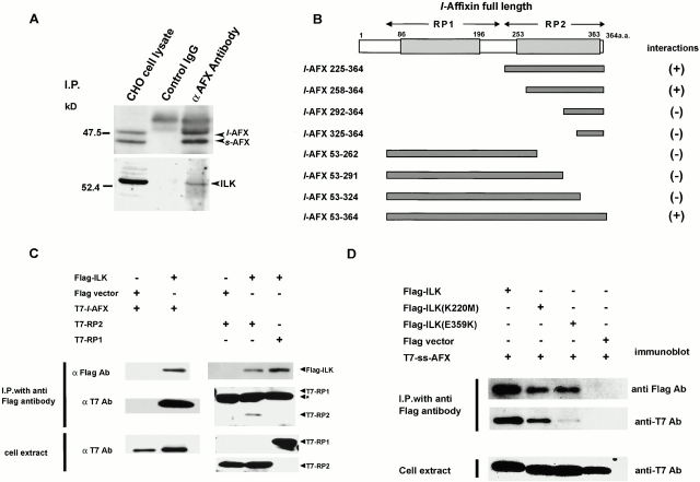
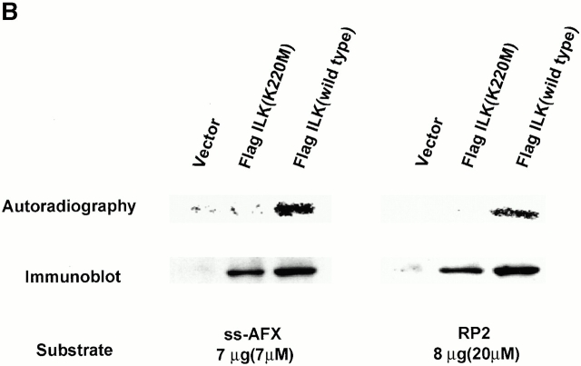
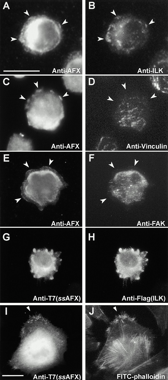
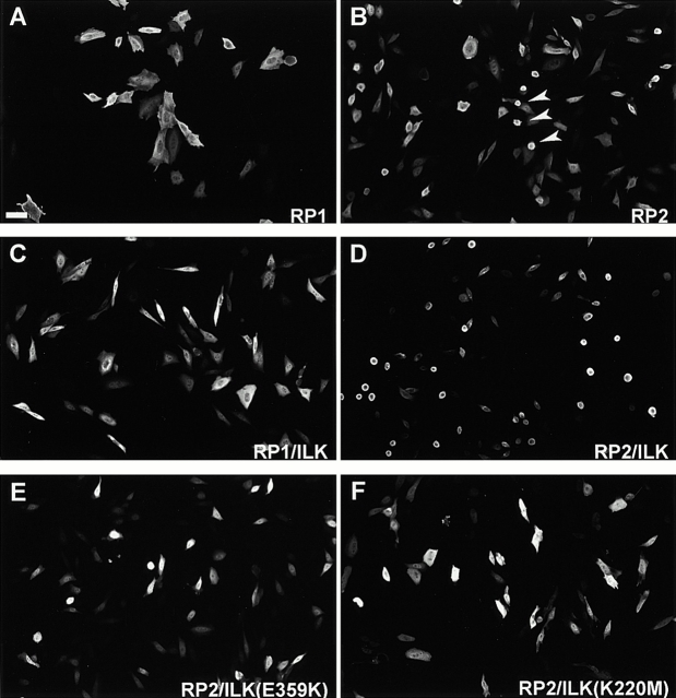
Focal adhesions (FAs) are essential structures for cell adhesion, migration, and morphogenesis. Integrin-linked kinase (ILK), which is capable of interacting with the cytoplasmic domain of beta1 integrin, seems to be a key component of FAs, but its exact role in cell-substrate interaction remains to be clarified. Here, we identified a novel ILK-binding protein, affixin, that consists of two tandem calponin homology domains. In CHOcells, affixin and ILK colocalize at FAs and at the tip of the leading edge, whereas in skeletal muscle cells they colocalize at the sarcolemma where cells attach to the basal lamina, showing a striped pattern corresponding to cytoplasmic Z-band striation. When CHO cells are replated on fibronectin, affixin and ILK but not FA kinase and vinculin concentrate at the cell surface in blebs during the early stages of cell spreading, which will grow into membrane ruffles on lamellipodia. Overexpression of the COOH-terminal region of affixin, which is phosphorylated by ILK in vitro, blocks cell spreading at the initial stage, presumably by interfering with the formation of FAs and stress fibers. The coexpression of ILK enhances this effect. These results provide evidence suggesting that affixin is involved in integrin-ILK signaling required for the establishment of cell-substrate adhesion.


Figures

Figure 1
Figure 1
Predicted amino acid sequence of human affixin. Predicted amino acid sequence of human affixin (sequence data available from GenBank/EMBL/DDBJ under accession no. AB048276) is aligned with the predicted affixin homologue in C. elegans (accession no. T21D12.4) and D. melanogaster (accession no. AAF49016) based on analysis using clustal W. Residues similar and identical to those of affixin are lightly and heavily shaded, respectively. The second and third alternative initiation methionines are indicated by an arrowhead and an asterisk, respectively. Two CH domains are underlined.
Figure 2
Figure 2
Affixin is a novel CH domain–containing protein. (A) Alignment of the two CH domains of human affixin (NH2-terminal CH and COOH-terminal CH) with the first CH domains in the actin-binding region of three human actin cross-linking proteins (spectrin β, sequence data available from GenBank/EMBL/DDBJ under accession no. M96803; α-actinin, accession no. P12814; dystrophin, accession no. P11532) and the single CH domain of human calponin H1 (accession no. SWP51911). Residues similar and identical to those of affixin are lightly and heavily shaded, respectively. Asterisks at the top indicate the consensus residues that are essentially conserved among CH domain–containing proteins (Carugo et al. 1997). The underlined sequence indicates the COOH-terminal hydrophobic residues conserved in the first CH domains of the actin-linking proteins. (B) Schematic diagram of the overall structure of the predicted affixin protein. Human affixin comprises two tandem CH domains, both of which show the closest similarity to the first CH domain in the actin-binding site of human β-spectrin. In contrast to spectrin, affixin is unique in that it lacks the triple-helical repeats (Sr, spectrin repeats). A C. elegans homologue of affixin (accession no. T21D12.4) is also shown at the bottom.
Figure 4
Figure 4
Detailed analysis of the affixin–ILK interaction. (A) Coimmunoprecipitation of ILK with endogenous affixin. Subconfluent CHO cells were lysed in immunoprecipitation buffer. Antiaffixin and control IgG immunoprecipitates were subjected to SDS-PAGE followed by Western blotting with antiaffixin (top) and ILK (bottom) antibodies. (B) Mapping of the ILK-binding region on affixin molecule by yeast two-hybrid binding assay. cDNA fragments encoding human l-affixin deletion mutants and full-length ILK were subcloned into pAS2-1 and pGAD424 vectors, respectively. These vectors were cotransformed into yeast Y187(a), and 5 d later the interactions were examined by β-galactosidase filter assay. (C) COOH-terminal half of affixin is coimmunoprecipitated with ILK from COS-7 cells. Expression vectors encoding the T7-tagged ss-affixin (left) or its NH2-terminal (RP1) or COOH-terminal (RP2) half (right) were cotransfected into COS-7 cells with pME18s-Flag expression vectors with or without a full-length ILK cDNA insert. The immunoprecipitation assay was performed using anti-Flag antibody, and immunocomplexes were subjected to immunoblot analysis with anti-Flag and anti-T7 antibody. The asterisk indicates a band corresponding to the IgG light chain. (D) The binding of T7-tagged ss-affixin with Flag-tagged ILK point mutants was examined by immunoprecipitation assays in COS-7 cells. Immunoprecipitation was performed with anti-Flag monoclonal antibody, whereas Western blot analysis of the immunocomplex was carried out using anti-T7 and anti-Flag antibody. ILK(E359K) and ILK(K220M) represent point mutants in which glutamate 359 in the activation loop or lysine 220 in the ATP-binding region of the ILK kinase domain is replaced with lysine or methionine, respectively. Note that K220M but not E359 coimmunoprecipitates with affixin.
Figure 3
Figure 3
Expression of affixin mRNA and protein in various tissues and cell lines. (A) Human multiple Northern blots (CLONTECH Laboratories, Inc.) on which 2 μg of polyA RNAs isolated from the indicated tissues were probed with an affixin cDNA fragment (206–481 bp). (B) Western blot analysis of total lysates of CHO-K1 (lane 4) and NIH3T3 (lane 5) cells probed with the antiaffixin antibody. In lanes 1–3, total lysates of COS-7 cells transfected with pMe18s vector alone (lane 1), s-affixin (lane 2), and l-affixin (lane3) are loaded. Note that s-affixin is also detected in lysate of cell transfected with an expression vector encoding l-affixin. (C) 15 μg of various tissue lysates from adult rat was loaded and analyzed with the antiaffixin antibody as indicated. Note that a strong band with a lower molecular weight was specifically detected in spleen. The migration rate of this band is the same as that of ss-affixin that is expressed using the third initiation methionine (data not shown; see Fig. 1). AFX, affixin.
Figure 5
Figure 5
ILK phosphorylates affixin in vitro. (A) Kinase activity of wild-type ILK and its point mutants overexpressed in COS-7 cells. Flag-tagged ILK or its mutants were overexpressed and immunoprecipitated from COS-7 cell lysates with monoclonal anti-Flag antibody. Kinase activities of the resultant immunocomplexes were examined using MBP as a substrate. Top, autoradiography showing 32P incorporation into MBP; bottom, Western blot analysis of the precipitated Flag-tagged ILK or its mutants using polyclonal anti-Flag antibody. (B) Full-length and COOH-terminal half of ss-affixin are phosphorylated by ILK in vitro. Recombinant full-length and COOH-terminal half of ss-affixin (RP2) were used as substrates to estimate the kinase activity of the ILK immunocomplexes prepared as described in A.
Figure 5
Figure 5
ILK phosphorylates affixin in vitro. (A) Kinase activity of wild-type ILK and its point mutants overexpressed in COS-7 cells. Flag-tagged ILK or its mutants were overexpressed and immunoprecipitated from COS-7 cell lysates with monoclonal anti-Flag antibody. Kinase activities of the resultant immunocomplexes were examined using MBP as a substrate. Top, autoradiography showing 32P incorporation into MBP; bottom, Western blot analysis of the precipitated Flag-tagged ILK or its mutants using polyclonal anti-Flag antibody. (B) Full-length and COOH-terminal half of ss-affixin are phosphorylated by ILK in vitro. Recombinant full-length and COOH-terminal half of ss-affixin (RP2) were used as substrates to estimate the kinase activity of the ILK immunocomplexes prepared as described in A.
Figure 6
Figure 6
Colocalization of affixin with ILK at FAs and the tip of the leading edge in CHO cells. Immunofluorescence staining with antiaffixin antibody of CHO cells (A and C) or with anti-T7 antibody of CHO cells transfected with T7-tagged affixin (E and G). Cells were stained simultaneously with antivinculin (B and F), anti-ILK (D) antibodies, or rhodamine-phalloidin (H). In E–H, CHO cells are reseeded on fibronectin-coated coverslips and fixed after 4-h spreading. Note that affixin and ILK are colocalized at FAs visualized by vinculin staining (A, B, and E and F, arrowheads) and at the tip of the leading edge (A–D, arrowheads). In C and D, fixation was performed with 100% cold methanol, whereas in other cases 1 (A and B) or 2% (E–H) paraformaldehyde in PBS was used. Intense signals from nuclei or perinuclear region represent nonspecific staining of the antiaffixin antibody observed depending on the fixation conditions. Bar, 25 μm.
Figure 7
Figure 7
Affixin and ILK localize at the sarcolemma of skeletal muscle cells. Immunofluorescence analysis of cross (A) or longitudinal (B and G) sections of human skeletal muscle with antiaffixin antibody. Cells were stained simultaneously with anti-ILK antibody (B and E) and anti–α-actinin antibody (H). Merged views are also demonstrated (C, F, and I). Observations were performed using confocal microscopy, and in D–I optical sections (0.4 μm) that tangentially cut off the sarcolemma were selected to demonstrate the striped pattern of affixin and ILK staining on the sarcolemma. In this section, staining of Z-bands with anti–α-actinin almost disappears, but in some regions the completely matched correlation between affixin staining and Z-band is clearly demonstrated. Note that affixin concentrated in the sites of the sarcolemma where Z-bands anchor (I, arrowheads). Bars, 25 μm.
Figure 9
Figure 9
Overexpression of the affixin deletion mutant, RP2, inhibits the progression of the cell-spreading process. (A) Western blot analysis showing relative expression levels of each construct in CHO-K1 cells. The total lysates of CHO-K1 transfected with indicated expression vectors were prepared 48 h after transfection and analyzed using anti-T7 antibody. (B) CHO cells transfected with T7-tagged ss-affixin or its deletion mutants (RP1 or RP2) were replated on fibronectin-coated coverslips 48 h after transfection. 1 or 4 h later, the cells were fixed and stained with anti-T7 polyclonal antibody. (C) The numbers of transfected cells with round morphology were counted at 1 and 4 h after replating. As control, CHO cells treated in a manner similar to the transfected cells were stained with rhodamine-phalloidin and counted. Note that cells transfected with RP2 but not ss-affixin or RP1 are blocked in the round morphology even at 4 h after reseeding. The values given represent mean values (± SD) of three independent experiments. (D) Cells overexpressing T7-tagged RP2 were doubly stained with anti-T7 polyclonal antibody (left) and FITC-phalloidin or antivinculin monoclonal antibody as indicated. Bars, 25 μm.
Figure 9
Figure 9
Overexpression of the affixin deletion mutant, RP2, inhibits the progression of the cell-spreading process. (A) Western blot analysis showing relative expression levels of each construct in CHO-K1 cells. The total lysates of CHO-K1 transfected with indicated expression vectors were prepared 48 h after transfection and analyzed using anti-T7 antibody. (B) CHO cells transfected with T7-tagged ss-affixin or its deletion mutants (RP1 or RP2) were replated on fibronectin-coated coverslips 48 h after transfection. 1 or 4 h later, the cells were fixed and stained with anti-T7 polyclonal antibody. (C) The numbers of transfected cells with round morphology were counted at 1 and 4 h after replating. As control, CHO cells treated in a manner similar to the transfected cells were stained with rhodamine-phalloidin and counted. Note that cells transfected with RP2 but not ss-affixin or RP1 are blocked in the round morphology even at 4 h after reseeding. The values given represent mean values (± SD) of three independent experiments. (D) Cells overexpressing T7-tagged RP2 were doubly stained with anti-T7 polyclonal antibody (left) and FITC-phalloidin or antivinculin monoclonal antibody as indicated. Bars, 25 μm.
Figure 8
Figure 8
Distribution of affixin during the early stages of the cell spreading process. CHO cells (A, C, and E) or those cotransfected with T7-tagged affixin and Flag-tagged ILK (G and I) were replated on fibronectin-coated coverslips and 1 h later fixed and stained with antiaffixin or anti-T7 antibodies as indicated. Cells were stained simultaneously with anti-ILK (B), antivinculin (D), anti-FAK (F), anti-Flag (H) antibodies, or FITC-phalloidin (J). Note that affixin is concentrated in peripheral blebs (A–F, arrowheads), whereas in well-spread cells dot-like staining is observed from which F-actin bundles emanate (I and J). Fixation is 100% methanol (A–F), 2% paraformaldehyde (G–J). Bars, 25 μm.
Figure 10
Figure 10
Overexpression of the COOH-terminal half of affixin in well-spread cells induces ILK-dependent disruption of cell–substrate interaction. T7-tagged affixin mutants (RP1 or RP2) were overexpressed alone (A and B), with Flag-tagged ILK (C and D), or with ILK point mutants (E and F) in CHO cells as indicated. 48 h after transfection, the cells were stained with anti-T7 polyclonal antibody. Note that overexpression of RP2 but not RP1 induces cell rounding, and this effect of RP2 is greatly enhanced by the coexpression of ILK but not ILK(E359) or ILK(K220M). Bar, 25 μm.

References

    1. Bauer J.S., Varner J., Schreiner C., Kornberg L., Nicholas R., Juliano R.L. Functional role of the cytoplasmic domain of the integrin alpha 5 subunit. J. Cell Biol. 1993;122:209–221. - PMC - PubMed
    1. Bereiter-Hahn J., Luck M., Miebach T., Stelzer H.K., Voth M. Spreading of trypsinized cellscytoskeletal dynamics and energy requirements. J. Cell Sci. 1990;96:171–188. - PubMed
    1. Carugo K.D., Banuelos S., Saraste M. Crystal structure of a calponin homology domain. Nat. Struct. Biol. 1997;4:175–179. - PubMed
    1. Cunningham C.C. Actin polymerization and intracellular solvent flow in cell surface blebbing. J. Cell Biol. 1995;129:1589–1599. - PMC - PubMed
    1. Dedhar S., Williams B., Hannigan G. Integrin-linked kinase (ILK)a regulator of integrin and growth-factor signalling. Trends Cell Biol. 1999;9:319–323. - PubMed

Publication types

MeSH terms