. 2001 Jun 11;153(6):1315-26.
doi: 10.1083/jcb.153.6.1315.

Localization of calmodulin and dynein light chain LC8 in flagellar radial spokes

Affiliations

Localization of calmodulin and dynein light chain LC8 in flagellar radial spokes

P Yang et al. J Cell Biol. .

Abstract

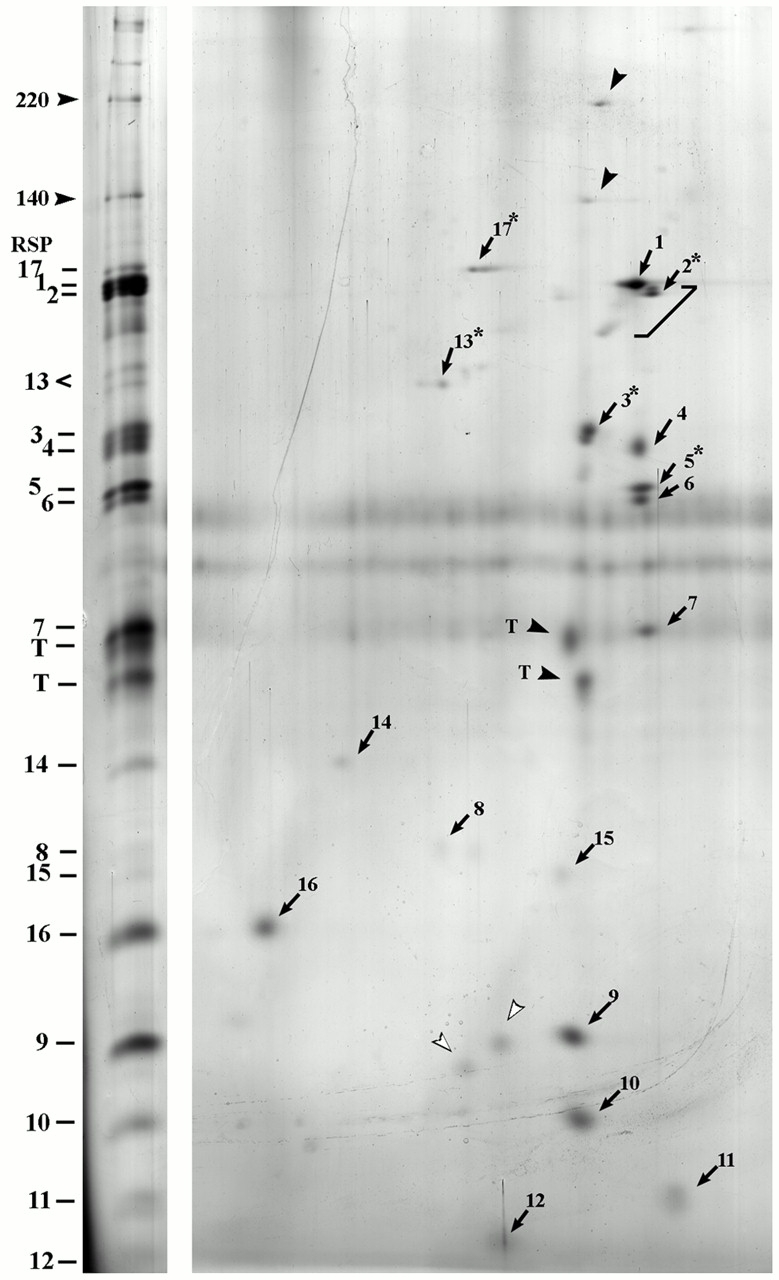
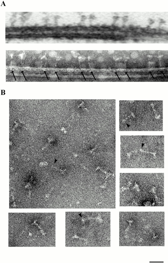
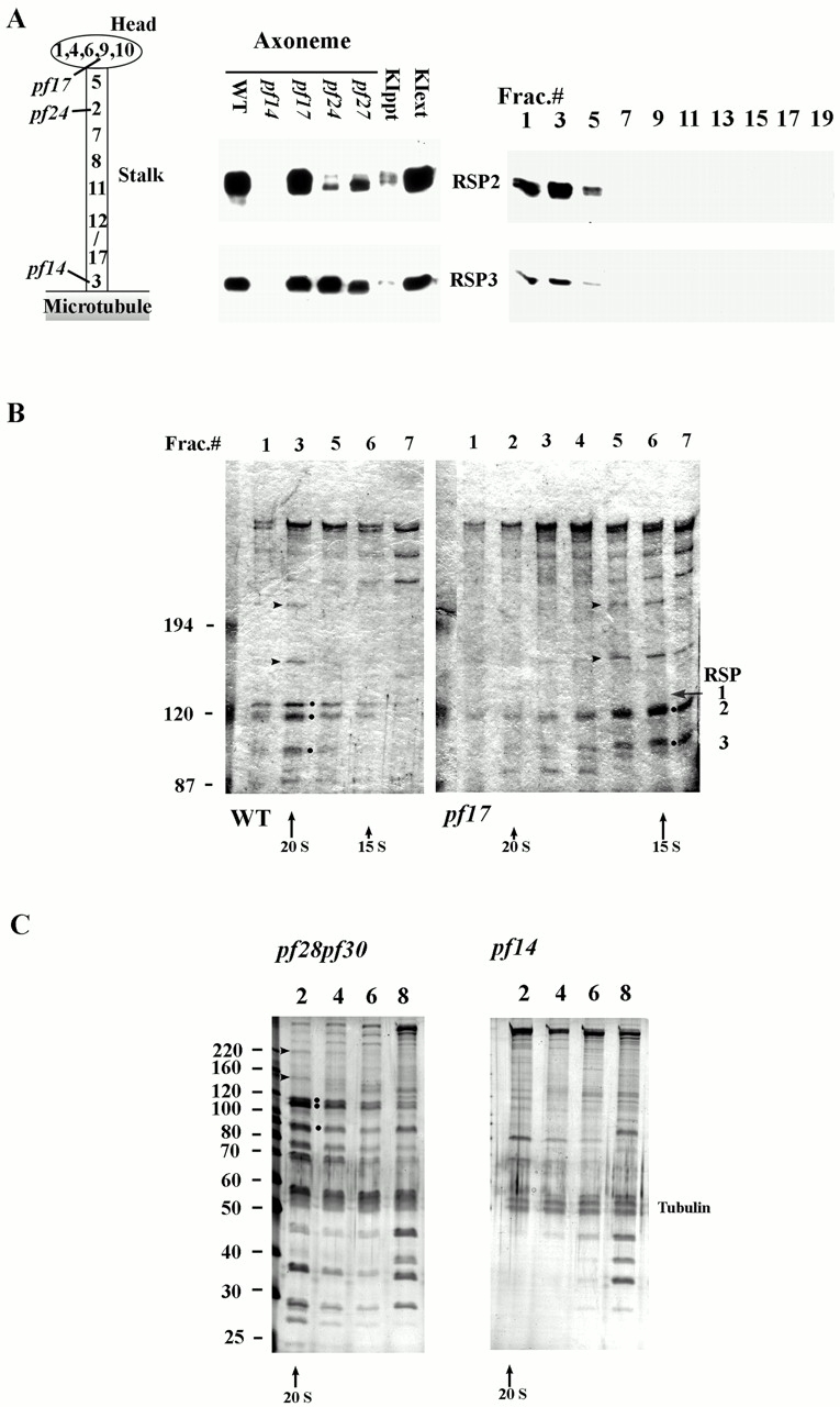
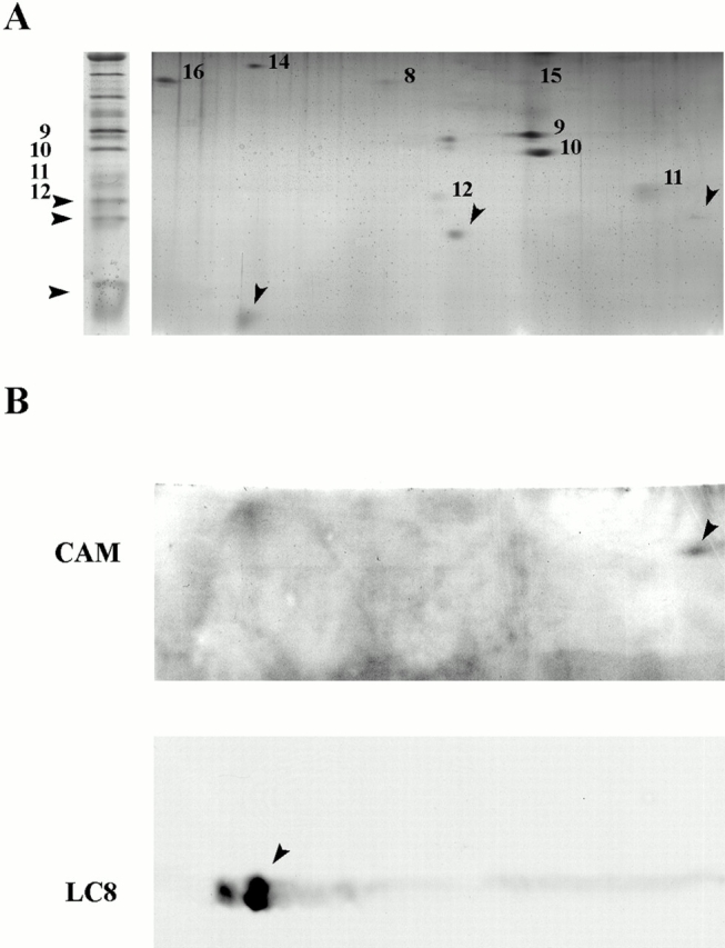
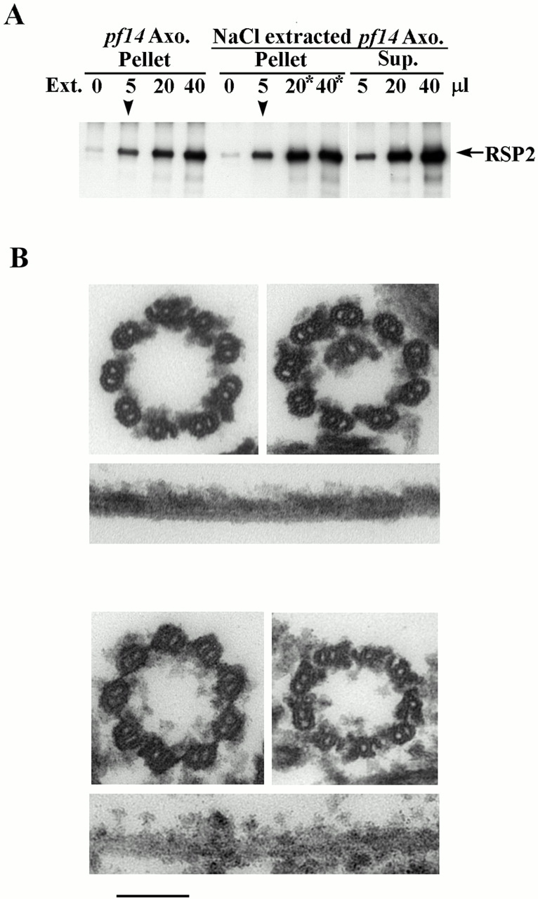
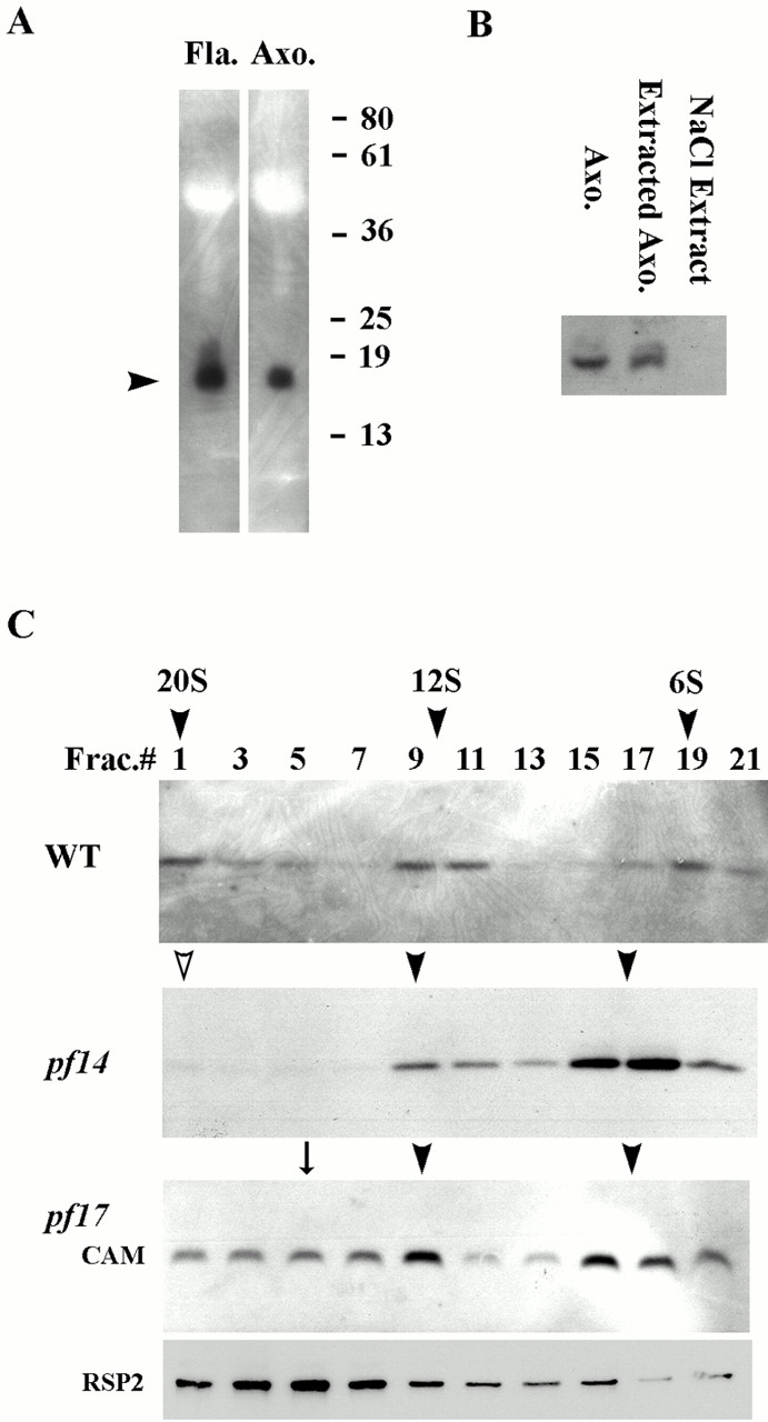
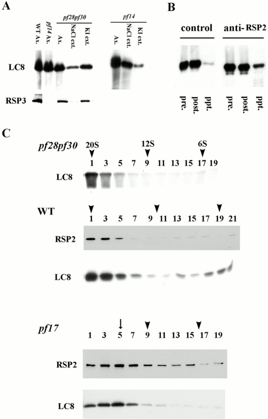
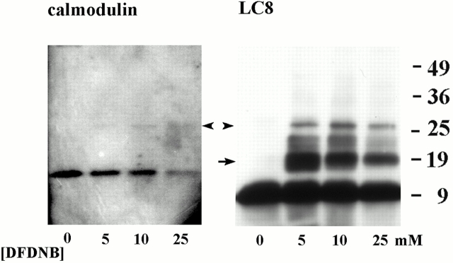
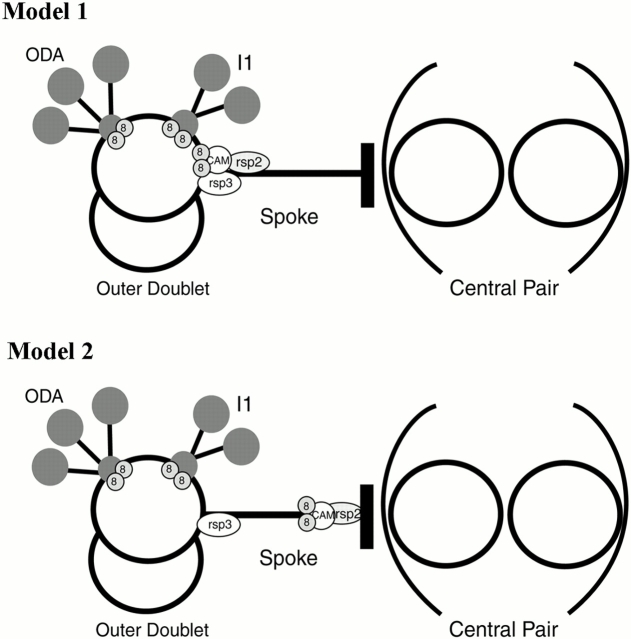
Genetic and in vitro analyses have revealed that radial spokes play a crucial role in regulation of ciliary and flagellar motility, including control of waveform. However, the mechanisms of regulation are not understood. Here, we developed a novel procedure to isolate intact radial spokes as a step toward understanding the mechanism by which these complexes regulate dynein activity. The isolated radial spokes sediment as 20S complexes that are the size and shape of radial spokes. Extracted radial spokes rescue radial spoke structure when reconstituted with isolated axonemes derived from the radial spoke mutant pf14. Isolated radial spokes are composed of the 17 previously defined spoke proteins as well as at least five additional proteins including calmodulin and the ubiquitous dynein light chain LC8. Analyses of flagellar mutants and chemical cross-linking studies demonstrated calmodulin and LC8 form a complex located in the radial spoke stalk. We postulate that calmodulin, located in the radial spoke stalk, plays a role in calcium control of flagellar bending.


Figures

Figure 2
Figure 2
The 20S radial spoke complex contains all 17 previously identified spoke proteins (compare to Piperno et al. 1981). Proteins in one-dimensional (left) and 2D (right) gels of the 20S fraction from pf28pf30 are revealed by silver staining. The 17 radial spoke proteins are numbered. In addition, present in the 20S fraction are the 140- and 210-kD proteins (arrowheads), as well as two unknown proteins (open arrowheads) and tubulin (T). Phosphorylated proteins are labeled (*), and RSP2 distribution, based on Western blots, are indicated by the bracket. Acidic polypeptides are on the right. No polypeptides are present in the basic half of the gel (not shown). A 9% slab gel was used for the second dimension.
Figure 3
Figure 3
The 20S radial spoke fraction contains T-shaped particles with the dimensions of intact radial spoke in situ. (A) Longitudinal images of thin section (top) and negatively stained (bottom) doublet microtubules, viewed edge-on, reveals paired radial spoke structures (arrows; compare with Witman et al. 1978). (B) Negative-stained images of the 20S radial spoke particles. A globular structure (arrowhead) was often found at the proximal end of the isolated spokes. Bar, 40 nm.
Figure 1
Figure 1
Extraction of flagellar radial spoke proteins and isolation of a 20S radial spoke complex and a 15S radial spoke stalk complex. (A, left) Diagram illustrating the predicted location of radial spoke proteins and the gene products of pf14, pf17, and pf24 in the radial spoke stalk and head (Curry and Rosenbaum 1993). (middle) Western blot analyses of isolated axonemes and axonemal fractions, using antibodies to RSP2 and -3 (lane 1, wild-type axonemes after extraction in 0.6 M NaCl; (lanes 2–5) axonemes from pf14, pf17, pf24, and pf27; (lane 6) wild-type axonemes after 0.6 M KI extraction; (lane 7) KI extract from wild-type axonemes. Notably, RSP2 and -3 are extracted with 0.6 M KI (middle, compare lanes 6 and 7) and cosediment at ∼20S in the 5–20% sucrose gradient (right, fraction 3). Protein loads and transfers were controlled by Ponceau red staining of blots (not shown). (right) Sucrose gradient fractions of the KI extract from wild-type cells probed with antibodies to RSP2 and -3. (B) Coomassie-stained 5% gels of sucrose gradient fractions reveal cosedimentation of several proteins in addition to RSP2 and -3. RSP1, -2, and -3 are indicated (•; left panel, fraction 3). Extraction of pf17 axonemes yields a diffuse 15S peak containing a subset of the proteins found in the 20S complex. Arrowheads indicate the 140- and 210-kD proteins that cosediment with both the 20S radial spoke and 15S radial spoke stalk complexes. RSP1 (gray arrow), a spoke head protein, is missing in the 15S complex. (C) Silver-stained 8% gels of sucrose gradient fractions of the axonemal extract from pf28pf30, a mutant lacking outer arm dynein and inner arm dynein I1. The 20S radial spoke complex is composed of several proteins (left), that are missing in the 20S fractions from a mutant lacking the radial spokes, pf14 (right panel). Samples analyzed in B and C were derived from 0.5 M KI (B) or 0.6 M NaBr (C) extract of axonemes first extracted in 0.6 M NaCl. Molecular weights are indicated on the left.
Figure 5
Figure 5
In addition to the radial spoke proteins described above, three small proteins including calmodulin, and dynein light chain LC8 are associated with the isolated radial spoke. (A) The 20S radial spoke fraction was separated by 14% one-dimensional (left) and 2D (right) gels. Silver staining revealed three proteins with masses of ∼18, ∼16, and ∼10 kD (arrowheads). (B) Western blot analyses revealed that the 18-kD protein is recognized by anti–Dictystelium calmodulin monoclonal antibody (top). The 10-kD protein is recognized by anti-LC8 antibody (bottom). The identity of the 16-kD protein is not known.
Figure 4
Figure 4
In vitro reconstitution of radial spokes. Western blots (A) and electron microscopy (B) reveal that extracted radial spokes bind to pf14 axonemes and restore radial spoke structure. Increasing amounts of extract containing radial spokes were incubated with either pf14 axonemes (lanes 1–4) or pf14 axonemes preextracted with 0.6 M NaCl (lanes 5–11). Pellets containing axonemes (lanes 1–4 for pf14 axonemes, and lanes 5–8 for NaCl extracted pf14 axonemes), and the supernatant for the NaCl-extracted pf14 axonemes (lane 9–11) were analyzed by Western analysis using RSP2 antibody. A 1:1 stoichiometry of radial spokes to the binding sites was predicted when 5 μl spoke fraction (arrowheads) was added to the axonemes. Rebinding saturated (*) when 20 μl extract was added to NaCl-extracted axonemes. (B) Cross and longitudinal sections of NaCl extracted pf14 axonemes (top), and the same axonemes reconstituted with extracted radial spokes (bottom). Notably, spoke structures are restored to the A-microtubules (cross section view). Bar, 96 nm.
Figure 6
Figure 6
Calmodulin is localized in the radial spoke stalk. (A) Western blot analyses revealed that a fraction of flagellar calmodulin tightly associates with axoneme. (B) The axonemal fraction is not extractable with 0.6 M NaCl. (C) On sucrose gradients, calmodulin in the KI extract sediments as three peaks when derived from wild-type axonemes (top, arrowheads). The 20S calmodulin peak is absent in extracts derived from pf14 (open arrowhead, middle). In the KI extract from pf17 axonemes, the discrete 20S calmodulin peak is missing, and rather it sediments as a diffuse peak (arrow, third panel) that overlaps with the second 12S calmodulin peak and cosediments with radial spoke stalk proteins including RSP2 (compare third and fourth panels, C).
Figure 7
Figure 7
Dynein light chain LC8 is a subunit of the radial spoke stalk, as well as a subunit of dynein structures. (A) Western blot analyses of LC8 and RSP3 in wild-type axonemes (lane 1); pf14 axonemes (lane 2); pf28pf30 axonemes (lane 3); 0.6 M NaCl extract (lane 4); and 0.6 M KI extract (lane 5). Notably, LC8 is abundant in pf14 and pf28pf30 axonemes, and in pf28pf30 axonemes, the majority of LC8, and RSP3 (bottom), resists extraction in 0.6 M NaCl, consistent with the localization in radial spokes. In contrast, LC8 in pf14 is largely extractable in 0.6 M NaCl (right), consistent with this fraction of LC8 being a subunit of dynein. (B) Coprecipitation of LC8 with radial spokes. RSP2 antibody was used to precipitate the 20S radial spoke complex from KI extracts (right). Western blots of LC8 showed that LC8 is present in the immunoprecipitate (lane 3, right). In contrast, little LC8 is precipitated in control experiments performed without addition of RSP2 antibody (left, and compare lane 3 of right and left panels). (lane 1 of each panel) Extract before precipitation (pre). (lane 2 of each panel) Extract after precipitation (post). (C) LC8 is located in the radial spoke stalk. Western analyses, using anti-LC8 and anti-RSP2, of sucrose gradient fractions from 0.5 M KI extracts from pf28pf30 (top) and wild type (middle). In each case, LC8 cosediments with the 20S radial spoke complex. In contrast, LC8 sediments as a diffuse peak centered at ∼15S (arrow, fraction 5, bottom) when derived from pf17 axonemes.
Figure 8
Figure 8
Cross-linking analysis reveals dimerization of LC8 and close interaction of calmodulin and LC8 in the radial spoke stalk. The 15S fraction derived from pf17axonemes was treated with DFDNB at concentrations indicated. Samples were then prepared for Western blot analyses using anticalmodulin (left) or anti-LC8 (right) antibodies. A 27-kD cross-linked product reacted with both antibodies (arrowheads). A 20-kD LC8 dimer also results from cross-linking (arrow, right).
Figure 9
Figure 9
Two models depicting the possible location of the key proteins in the radial spoke stalk including, LC8, calmodulin, RSP2, and RSP3.

Similar articles

Cited by

References

    1. Bannai H., Yoshima M., Takahashi K., Shingyoji C. Calcium regulation of microtubule sliding in reactivated sea urchin sperm flagella. J. Cell Sci. 2000;113:831–839. - PubMed
    1. Benashski S.E., Harrison A., Patel-King R.S., King S.M. Dimerization of the highly conserved light chain shared by dynein and myosin V. J. Biol. Chem. 1997;272:20929–20935. - PubMed
    1. Bessen M., Fay R.B., Witman G.B. Calcium control of waveform in isolated flagellar axonemes of Chlamydomonas . J. Cell Biol. 1980;86:446–455. - PMC - PubMed
    1. Brokaw C.J. Regulation of sperm flagellar motility by calcium and cAMP-dependent phosphorylation. J. Cell. Biochem. 1987;35:175–184. - PubMed
    1. Brokaw C.J. Control of flagellar bendinga new agenda based on dynein diversity. Cell Motil. Cytoskel. 1994;28:199–204. - PubMed

Publication types