. 2001 Jun;13(6):1369-82.
doi: 10.1105/tpc.13.6.1369.

Nodule-specific regulation of phosphatidylinositol transfer protein expression in Lotus japonicus

Affiliations

Nodule-specific regulation of phosphatidylinositol transfer protein expression in Lotus japonicus

P Kapranov et al. Plant Cell. 2001 Jun.

Abstract

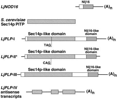
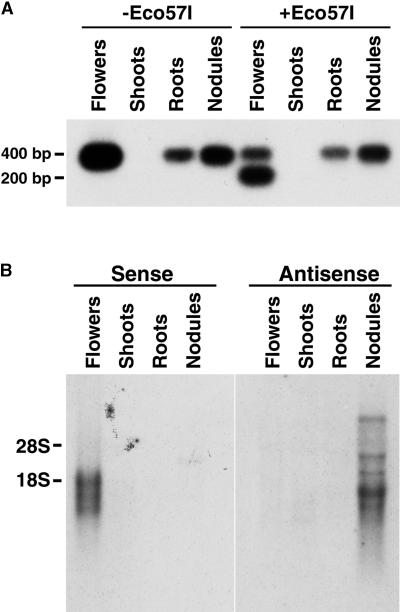
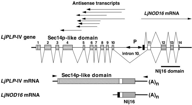
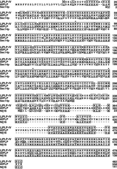
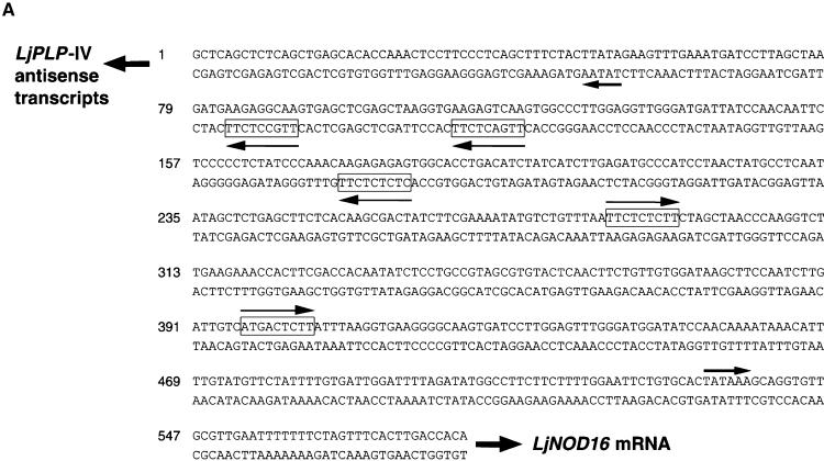
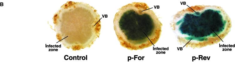
Phosphatidylinositol transfer proteins (PITPs) modulate signal transduction pathways and membrane-trafficking functions in eukaryotes. Here, we describe the characterization of a gene family from Lotus japonicus that encodes a novel class of plant PITP-like proteins (LjPLPs) and that is regulated in an unusual nodule-specific manner. Members of this gene family were identified based on their nucleotide sequence homology with a previously described cDNA, LjNOD16, which encodes the L. japonicus late nodulin Nlj16. Nlj16 or highly related amino acid sequences are shown to constitute C-terminal domains of LjPLPs and are suggested to function as specific plasma membrane targeting modules. The expression patterns of one member of this gene family (LjPLP-IV) revealed that LjNOD16 mRNA synthesis in nodules is the result of the transcriptional activity of a nodule-specific promoter located in an intron of the LjPLP-IV gene. This intron-borne bidirectional promoter also generates nodule-specific antisense transcripts derived from the N-terminal PITP domain coding region of the LjPLP-IV gene. We propose that Nlj16 protein synthesis and LjPLP-IV antisense transcript generation are components of an elaborate mechanism designed to control LjPLP synthesis and/or functioning in nodules.


Figures

Figure 1.
Figure 1.
Scheme of LjPLP cDNAs. The boxes represent coding regions, and the lines correspond to the 5′ and 3′ untranslated regions (UTRs) of the cDNAs. The hatched boxes represent either Sec14p or Sec14p-like domains, the shaded boxes represent Nlj16-like domains, and the open boxes correspond to regions sharing no apparent sequence similarity with the other cDNAs. The positions of the stop TAG222 codon in LjPLP-I and the corresponding CAG222 codon in LjPLP-II are indicated. The asterisk indicates that the LjPLP-II cDNA sequence was deduced from the nucleotide sequence of the corresponding L. japonicus genomic region. The ORFs present in the LjPLP-IV antisense transcripts, which encode Sec14p-like domains, are represented by inversely hatched boxes, and the intron sequences are represented by lines. The nucleotide sequences for LjPLP-I, LjPLP-II, and LjPLP-III have GenBank accession numbers AF366900, AF366901, and AF367433, respectively. The nucleotide sequence of the LjPLP-IV gene, from which the LjPLP-IV antisense transcripts originate, has GenBank accession number AF367434.
Figure 2.
Figure 2.
Tissue-Specific Expression of the LjPLP-I, LjPLP-II, and LjPLP-IV Genes. (A) Fragments (400-bp), encompassing either the UAG222 stop codon or the CAG222 triplet-containing regions of LjPLP-I and LjPLP-II mRNAs, respectively, were amplified using RT-PCR. The 400- and 200-bp fragments observed in the samples digested with Eco57I correspond to the LjPLP-I and LjPLP-II transcripts, respectively. (B) Tissue-specific expression of the LjPLP-IV transcripts. Ten micrograms of total RNA was hybridized with radiolabeled, strand-specific, RNA probes complementary to the presumed sense or antisense transcripts of the LjPLP-IV gene. The positions of 28S and 18S rRNAs are indicated.
Figure 3.
Figure 3.
Schemes of the LjPLP-IV Genomic Region and the LjPLP-IV Sense Transcripts and the LjNOD16 Transcript. Exons are numbered and represented by boxes, introns are symbolized by thin lines, and thick lines indicate the 5′ and 3′ UTRs. Differently shaded boxes correspond to either Sec14p-like or Nlj16 domains, as described in the legend to Figure 1. The solid box represents the first exon of the LjNOD16 transcript. The positions of various cDNA clones corresponding to either LjPLP-IV antisense transcripts or the LjNOD16 transcript are indicated by arrows above the diagram of the LjPLP-IV gene. The localization of the bidirectional promoter (P) within intron 10 is indicated. The positions of primers used to amplify the LjPLP-IV mRNA are indicated by arrowheads.
Figure 4.
Figure 4.
Amino Acid Sequence Alignment between L. japonicus LjPLP-IV, Nodulin Nlj16, AtPLP (Hypothetical Arabidopsis Protein CAB16843.1), and Yeast Sec14p Proteins. The alignment was generated using the PileUp function of the Genetics Computer Group package (Madison, WI) and SeqVu software version 1.0.1 (Garvan Institute of Medical Research, Sydney, Australia). Similar residues are boxed, and identical amino acids are boxed and shaded. Dashes represent gaps in the sequences, and asterisks indicate the conserved amino acid residues required for PtdIns binding activity of the yeast Sec14p protein (Sha et al., 1998).
Figure 5.
Figure 5.
The Internal Bidirectional Promoter of the LjPLP-IV Gene Directs the Expression of the β-Glucuronidase Reporter Gene to the Central Infected Zone of L. corniculatus Nodules. (A) Nucleotide sequence of the intron-localized bidirectional promoter of the LjPLP-IV gene. The directions of transcription of the LjPLP-IV antisense transcripts and the LjNOD16 transcript are indicated by arrows. The positions and presumed orientations of TATA box–like sequences are indicated by arrows and nodulin gene conserved promoter motifs are indicated by boxed sequences. (B) Histochemical localization of GUS activity in nodule hand sections derived from transgenic L. corniculatus plants harboring the promoter–GUS fusions. VB, nodule vascular bundle (for further details see text).
Figure 5.
Figure 5.
The Internal Bidirectional Promoter of the LjPLP-IV Gene Directs the Expression of the β-Glucuronidase Reporter Gene to the Central Infected Zone of L. corniculatus Nodules. (A) Nucleotide sequence of the intron-localized bidirectional promoter of the LjPLP-IV gene. The directions of transcription of the LjPLP-IV antisense transcripts and the LjNOD16 transcript are indicated by arrows. The positions and presumed orientations of TATA box–like sequences are indicated by arrows and nodulin gene conserved promoter motifs are indicated by boxed sequences. (B) Histochemical localization of GUS activity in nodule hand sections derived from transgenic L. corniculatus plants harboring the promoter–GUS fusions. VB, nodule vascular bundle (for further details see text).
Figure 6.
Figure 6.
The Conserved Nlj16 Domain Mediates the Plasma Membrane Targeting of the LjPLP-IV Protein. (A) to (E) Subcellular localization of the mGFP5–Nlj16 fusion proteins in onion epidermal cells. Fluorescent images of onion cells expressing mGFP5 alone (A), the mGFP5–Nlj16 fusion ([B] and [D]), and the mGFP5–Nlj16ΔCC fusion (C) are shown. (E) is a bright-field image of the cell shown in (D). The onion cell shown in (D) and (E) has undergone plasmolysis, and mGFP5 fluorescence can be seen on the periphery of the protoplast. P, protoplast; V, vacuole; CW, cell wall. (F) Alignment of the C-terminal Nlj16-like domains of LjPLP-II, LjPLP-III, LjPLP-IV, and Arabidopsis AtPLP. Similar residues are boxed, and identical amino acids are boxed and shaded. Dashes represent gaps in the sequences.
Figure 6.
Figure 6.
The Conserved Nlj16 Domain Mediates the Plasma Membrane Targeting of the LjPLP-IV Protein. (A) to (E) Subcellular localization of the mGFP5–Nlj16 fusion proteins in onion epidermal cells. Fluorescent images of onion cells expressing mGFP5 alone (A), the mGFP5–Nlj16 fusion ([B] and [D]), and the mGFP5–Nlj16ΔCC fusion (C) are shown. (E) is a bright-field image of the cell shown in (D). The onion cell shown in (D) and (E) has undergone plasmolysis, and mGFP5 fluorescence can be seen on the periphery of the protoplast. P, protoplast; V, vacuole; CW, cell wall. (F) Alignment of the C-terminal Nlj16-like domains of LjPLP-II, LjPLP-III, LjPLP-IV, and Arabidopsis AtPLP. Similar residues are boxed, and identical amino acids are boxed and shaded. Dashes represent gaps in the sequences.
Figure 7.
Figure 7.
The LjPLP-IV Protein Complements the Temperature-Sensitive Phenotype of the Yeast sec14 Mutant and Exhibits PtdIns and PtdCho Transfer Activity. (A) The growth phenotype of yeast strain CTY1079 (sec14-1ts Δspo14) harboring the indicated constructs (see Methods). Individual transformants were streaked on selective (−Ura) media and grown at permissive (28°C) and nonpermissive (37°C) temperatures. (B) and (C) Phospholipid transfer activities. The ability of the recombinant LjPLP-IV Sec14p domain (LjPLP-IV–Sec14p) to transfer 3H-PtdIns (B) or 14C-PtdCho (C) was measured in cytosol prepared from E. coli. Activities are represented as the percentage of total input 3H-PtdIns or 14C-PtdCho transferred from donor membranes to unlabeled acceptor membranes during a 30-min incubation at 37°C. Cytosol values are presented as amounts of protein added to the assay. Closed circles, Sec14p; open squares, LjPLP-IV–Sec14p; open circles, E. coli cytosol. Assay blanks represented the addition of buffer alone to the transfer assay reactions. These reactions defined assay background, and background values were subtracted from each assay to yield transfer values. Ranges of 12,695 to 15,121 cpm and 22,697 to 28,560 cpm of input 3H-PtdIns and 14C-PtdCho, respectively, were used in each assay. Background values for these assays ranged from 237 to 374 for the PtdIns transfer assays and from 238 to 391 for the PtdCho transfer assays. The data shown are from a representative experiment of at least three independent assays.

Similar articles

Cited by

References

    1. Aitken, J.F., van Heusden, G.P.H., Temkin, M., and Dowhan, W. (1990). The gene encoding the phosphatidylinositol transfer protein is essential for cell growth. J. Biol. Chem. 265, 4711–4717. - PubMed
    1. Bankaitis, V.A., Malehorn, D.E., Emr, S.D., and Greene, R. (1989). The Saccharomyces cerevisiae SEC14 gene encodes a cytosolic factor that is required for transport of secretory proteins from the yeast Golgi complex. J. Cell Biol. 108, 1271–1281. - PMC - PubMed
    1. Bankaitis, V.A., Aitken, J.F., Cleves, A.E., and Dowhan, W. (1990). An essential role for a phospholipid transfer protein in yeast Golgi function. Nature 347, 561–562. - PubMed
    1. Cleves, A.E., McGee, T.P., and Bankaitis, V.A. (1991). Phospholipid transfer proteins: A biological debut. Trends Cell Biol. 1, 30–34. - PubMed
    1. Cunningham, E., Tan, S.W., Swigart, P., Hsuan, J., Bankaitis, V., and Cockcroft, S. (1996). The yeast and mammalian isoforms of phosphatidylinositol transfer protein can all restore phospholipase C–mediated inositol lipid signaling in cytosol-depleted RBL-2H3 and HL60 cells. Proc. Natl. Acad. Sci. USA 93, 6589–6593. - PMC - PubMed

Publication types

MeSH terms

LinkOut - more resources