. 2001 Jun;13(6):1383-400.
doi: 10.1105/tpc.13.6.1383.

Molecular characterization of functional domains in the protein kinase SOS2 that is required for plant salt tolerance

Affiliations

Molecular characterization of functional domains in the protein kinase SOS2 that is required for plant salt tolerance

Y Guo et al. Plant Cell. 2001 Jun.

Abstract

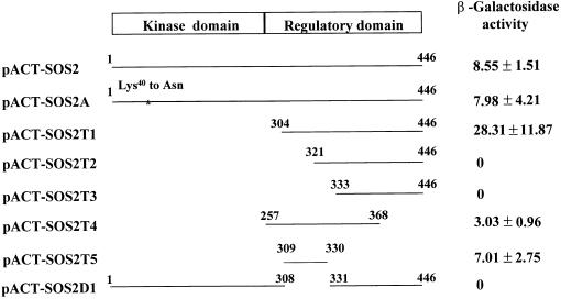
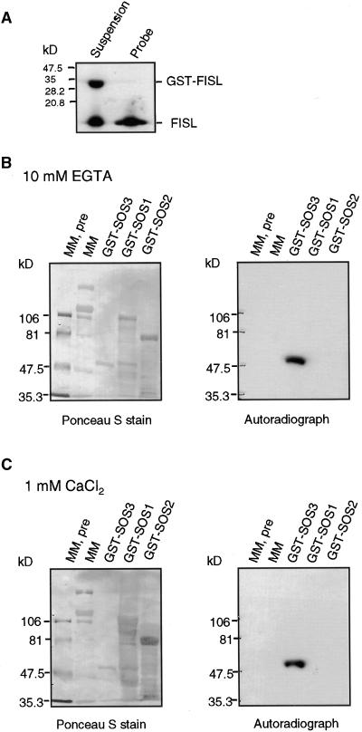
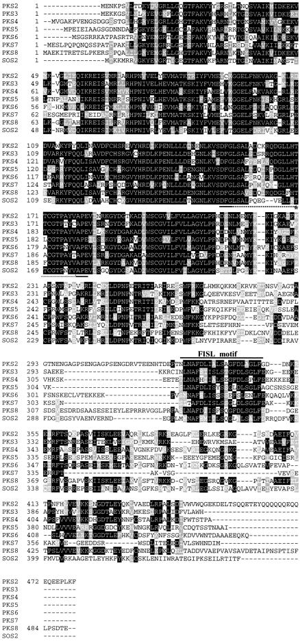
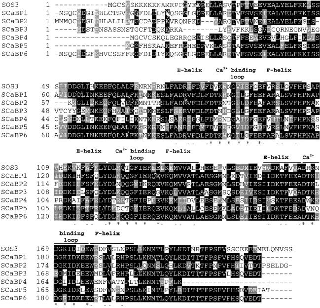
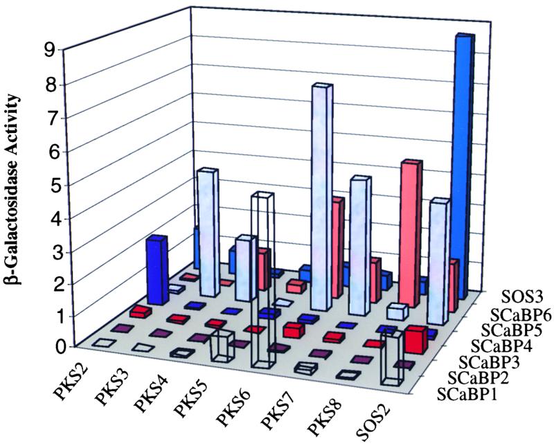
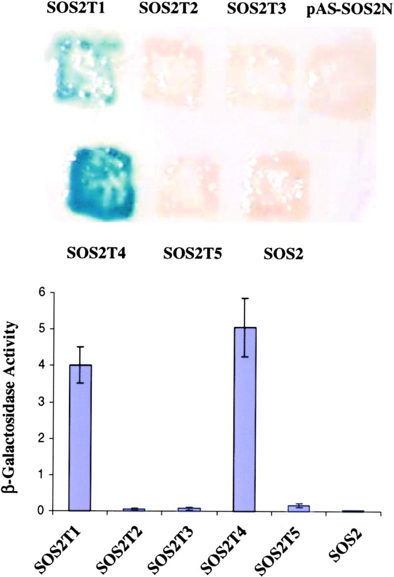
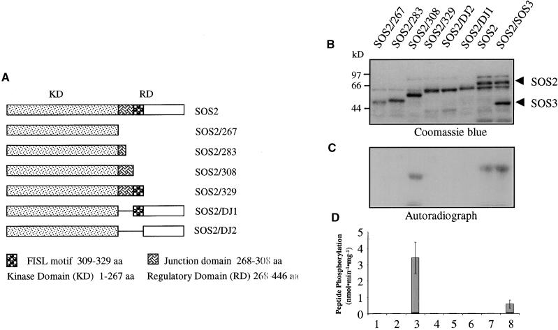
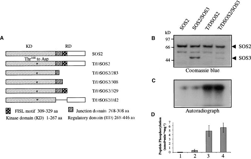
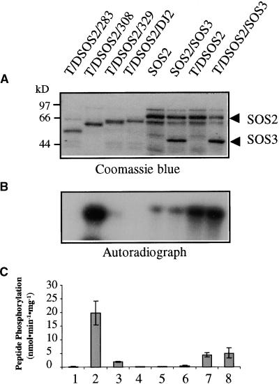
The SOS3 (for SALT OVERLY SENSITIVE3) calcium binding protein and SOS2 protein kinase are required for sodium and potassium ion homeostasis and salt tolerance in Arabidopsis. We have shown previously that SOS3 interacts with and activates the SOS2 protein kinase. We report here the identification of a SOS3 binding motif in SOS2 that also serves as the kinase autoinhibitory domain. Yeast two-hybrid assays as well as in vitro binding assays revealed a 21-amino acid motif in the regulatory domain of SOS2 that is necessary and sufficient for interaction with SOS3. Database searches revealed a large family of SOS2-like protein kinases containing such a SOS3 binding motif. Using a yeast two-hybrid system, we show that these SOS2-like kinases interact with members of the SOS3 family of calcium binding proteins. Two-hybrid assays also revealed interaction between the N-terminal kinase domain and the C-terminal regulatory domain within SOS2, suggesting that the regulatory domain may inhibit kinase activity by blocking substrate access to the catalytic site. Removal of the regulatory domain of SOS2, including the SOS3 binding motif, resulted in constitutive activation of the protein kinase, indicating that the SOS3 binding motif can serve as a kinase autoinhibitory domain. Constitutively active SOS2 that is SOS3 independent also was produced by changing Thr(168) to Asp in the activation loop of the SOS2 kinase domain. Combining the Thr(168)-to-Asp mutation with the autoinhibitory domain deletion created a superactive SOS2 kinase. These results provide insights into regulation of the kinase activities of SOS2 and the SOS2 family of protein kinases.


Figures

Figure 1.
Figure 1.
Deletion Analysis of SOS2 to Identify an SOS3 Binding Motif. Depicted are yeast two-hybrid constructs of SOS2 kinase with various deletions that were tested in combination with bait pAS-SOS3 in yeast for LacZ activation and quantified as β-galactosidase activity (n = 3). The C-terminal regulatory domain has been shown previously to be sufficient for binding to SOS3, whereas the N-terminal kinase catalytic domain is not required (Halfter et al., 2000). N-terminal deletion of 320 or 332 amino acids results in the loss of β-galactosidase activity, whereas the N-terminal deletion of 303 amino acids leads to very strong interaction. Amino acid residues from Met309 to Arg330 are sufficient and necessary to activate the LacZ reporter and are at a level comparable to that of the intact kinase protein.
Figure 2.
Figure 2.
The FISL Motif Binds to SOS3 in Vitro in Gel Blot Overlay Assays. (A) The 21–amino acid FISL motif identified in the yeast two-hybrid assay was produced as GST-FISL. GST-FISL was labeled with phosphorus-32 and, after cleavage with thrombin, which removed the GST portion, yielded 32P-FISL to be used as a probe. (B) GST-SOS3 and control proteins. Molecular mass markers, both prestained (MM, pre) and unstained (MM), GST-SOS3, GST-SOS1, and GST-SOS2 were separated on a 7.5% SDS-PAGE gel, electroblotted onto a membrane, and stained with Ponceau S to reveal all proteins (left). The membrane was overlaid with 32P-FISL in the presence of 10 mM EGTA and subjected to autoradiography (right). (C) As given in (B), except that the hybridization solution included 1 mM free Ca2+.
Figure 3.
Figure 3.
Amino Acid Alignment of SOS2 with PKS2 to PKS8. The N-terminal kinase catalytic domain is highly conserved. The C-terminal regulatory domain contains the conserved FISL motif (marked). Also marked is the activation loop between the conserved DFG and APE motifs (dots) and the Thr residue, which may be phosphorylated by an upstream protein kinase (asterisk). Dashed lines represent spaces that were introduced to maximize alignment.
Figure 4.
Figure 4.
Expression of PKS2 to PKS8 in Arabidopsis Shoots and Roots in Response to Salt Stress. Two-week-old Arabidopsis seedlings, grown on Murashige and Skoog (1962) nutrient agar plates, were treated with 100 or 200 mM NaCl and harvested at the specified times for RNA isolation. Twenty micrograms of poly(A)+ RNA was analyzed by RNA gel blotting. Ethidium bromide–stained rRNA is shown as a loading control. The blot was hybridized with gene-specific probes for PKS2 to PKS8.
Figure 5.
Figure 5.
Alignment of SOS3 with SCaBPs. Database searches revealed sequences similar to SOS3, that is, SCaBP1 to SCaBP6. All contain three EF-hand calcium binding motifs. The calcium binding loops, flanked by E and F helices, are marked. Asterisks and dashes denote amino acid residues important for calcium binding and the EF-hand structure, respectively (Moncrief et al., 1990).
Figure 6.
Figure 6.
β-Galactosidase Activity of Various Combinations between PKS Proteins and SCaBPs in a Yeast Two-Hybrid Assay. Yeast strain Y190 harboring SOS2 or PKS2 to PKS8 protein kinases in the pACT prey vectors were transformed with SOS3 or SCaBP1 to SCaBP6 in the bait vector pAS2. Their ability to activate the LacZ reporter gene, measured as β-galactosidase activity, was assayed as an indicator of the strength of their interaction. The activity values are the averages of three experiments. Standard deviations are not shown but are all within 5% of the respective average values. Numbers at left represent arbitrarily defined unit U.
Figure 7.
Figure 7.
Interaction between the Kinase and Regulatory Domains within the SOS2 Protein. Wild-type SOS2 and its deletion constructs depicted in Figure 1 were transformed into yeast strain Y190 harboring pAS-SOS2N as bait. Shown are β-galactosidase assays on a filter (top) or in liquid culture (bottom) for quantification of relative interaction strength. Numbers at left represent arbitrarily defined unit U. Error bars indicate ±sd.
Figure 8.
Figure 8.
Identification of an Autoinhibitory Domain in SOS2 by Deletion Analysis. C-terminal serial truncations or internal deletions of SOS2 were expressed in bacteria as GST fusion proteins and tested for autophosphorylation and phosphorylation of the peptide substrate p3. This analysis revealed that the SOS3-binding FISL motif can serve as the kinase autoinhibitory domain. (A) Diagram of SOS2 mutant constructs. aa, amino acids. (B) Coomassie blue–stained SDS-PAGE gel containing the SOS2 mutant proteins fused with GST. (C) Autoradiograph of the gel shown in (B) to detect kinase autophosphorylation. (D) Peptide phosphorylation activities of the proteins shown in (B). Error bars indicate ±sd (n = 3).
Figure 9.
Figure 9.
Activation of SOS2 Kinase Activity by the Thr168-to-Asp Mutation in the Activation Loop. (A) Diagram of SOS2 mutants that were expressed in bacteria as GST fusion proteins. aa, amino acids. (B) Coomassie blue–stained SDS-PAGE gel containing the relevant proteins. (C) Kinase autophosphorylation. (D) Phosphorylation of p3 by the kinases. Error bars indicate ±sd (n = 3).
Figure 10.
Figure 10.
Creation of a Superactive SOS2 Kinase by Combining the Thr168-to-Asp Mutation with the Autoinhibitory Domain Deletion. Construction of the SOS2 mutant proteins was as shown in Figure 9A. (A) Coomassie blue–stained SDS-PAGE gel containing the relevant proteins. (B) Kinase autophosphorylation. (C) Phosphorylation of p3 by the kinases. Error bars indicate ±sd (n = 3).
Figure 11.
Figure 11.
Effect of Ca2+ and Mg2+ on the Superactive Mutant SOS2 Kinase. (A) Coomassie blue–stained SDS-PAGE gel showing the mutant kinase protein (T/DSOS2/308). (B) Autophosphorylation activity of the kinase. (C) p3 phosphorylation activity of the kinase. (D) to (F) Same as (A) to (C), respectively, except for the indicated differences in Ca2+ and Mg2+ concentrations in the kinase reactions. Error bars in (C) and (F) indicate ±sd (n = 3).

References

    1. Arabidopsis Genome Initiative. (2000). Analysis of the genome sequence of the flowering plant Arabidopsis thaliana. Nature 408, 796–815. - PubMed
    1. Bush, D.S. (1995). Calcium regulation in plant cells and its role in signaling. Annu. Rev. Plant Physiol. Plant Mol. Biol. 46, 95–122.
    1. Celio, M.R., Pauls, T., and Schwaller, B. (1996). Guidebook to the Calcium-Binding Proteins. (Oxford, UK: Oxford University Press).
    1. Cohen, P., and Klee, C.B. (1988). Calmodulin. (New York: Elsevier).
    1. Cox, J.A. (1990). The role of intracellular calcium binding proteins. In Stimulus Response Coupling, J.-R. Dedman and V.L. Smith, eds. (Boston: CRC Press), pp. 266–269.

Publication types

MeSH terms