

Directed proteomics identifies a plant-specific protein rapidly phosphorylated in response to bacterial and fungal elicitors
- PMID: 11402173
- PMCID: PMC135571
- DOI: 10.1105/tpc.13.6.1467
Directed proteomics identifies a plant-specific protein rapidly phosphorylated in response to bacterial and fungal elicitors
Abstract
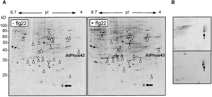
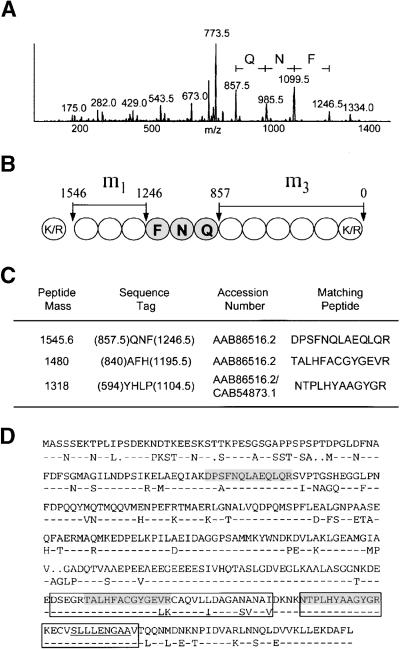
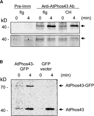
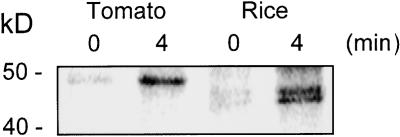
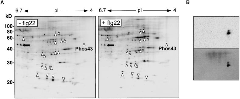
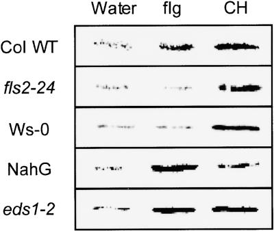
The perception of microbial signal molecules is part of the strategy evolved by plants to survive attacks by potential pathogens. To gain a more complete understanding of the early signaling events involved in these responses, we used radioactive orthophosphate to pulse-label suspension-cultured cells of Arabidopsis in conjunction with two-dimensional gel electrophoresis and mass spectrometry to identify proteins that are phosphorylated rapidly in response to bacterial and fungal elicitors. One of these proteins, AtPhos43, and related proteins in tomato and rice, are phosphorylated within minutes after treatment with flagellin or chitin fragments. By measuring (32)P incorporation into AtPhos43 immunoprecipitated from extracts of elicitor-treated hormone and defense-response mutants, we found that phosphorylation of AtPhos43 after flagellin treatment but not chitin treatment is dependent on FLS2, a receptor-like kinase involved in flagellin perception. Induction by both elicitors is not dependent on salicylic acid or EDS1, a putative lipase involved in defense signaling.
Figures
Similar articles
-
A plasma membrane syntaxin is phosphorylated in response to the bacterial elicitor flagellin.J Biol Chem. 2003 Nov 14;278(46):45248-54. doi: 10.1074/jbc.M307443200. Epub 2003 Aug 28. J Biol Chem. 2003. PMID: 12949074
-
The Arabidopsis receptor kinase FLS2 binds flg22 and determines the specificity of flagellin perception.Plant Cell. 2006 Feb;18(2):465-76. doi: 10.1105/tpc.105.036574. Epub 2005 Dec 23. Plant Cell. 2006. PMID: 16377758 Free PMC article.
-
Molecular identification and characterization of the tomato flagellin receptor LeFLS2, an orthologue of Arabidopsis FLS2 exhibiting characteristically different perception specificities.Plant Mol Biol. 2007 Jul;64(5):539-47. doi: 10.1007/s11103-007-9173-8. Epub 2007 May 25. Plant Mol Biol. 2007. PMID: 17530419
-
Flagellin perception: a paradigm for innate immunity.Trends Plant Sci. 2002 Jun;7(6):251-6. doi: 10.1016/s1360-1385(02)02261-6. Trends Plant Sci. 2002. PMID: 12049921 Review.
-
Defense Against Pathogens: Structural Insights into the Mechanism of Chitin Induced Activation of Innate Immunity.Curr Med Chem. 2017 Nov 24;24(36):3980-3986. doi: 10.2174/0929867323666161221124345. Curr Med Chem. 2017. PMID: 28003004 Review.
Cited by
-
Role of Protein Tyrosine Phosphatases in Plants.Curr Genomics. 2015 Aug;16(4):224-36. doi: 10.2174/1389202916666150424234300. Curr Genomics. 2015. PMID: 26962298 Free PMC article.
-
The receptor-like kinase SERK3/BAK1 is a central regulator of innate immunity in plants.Proc Natl Acad Sci U S A. 2007 Jul 17;104(29):12217-22. doi: 10.1073/pnas.0705306104. Epub 2007 Jul 11. Proc Natl Acad Sci U S A. 2007. PMID: 17626179 Free PMC article.
-
Flower proteome: changes in protein spectrum during the advanced stages of rose petal development.Planta. 2005 Sep;222(1):37-46. doi: 10.1007/s00425-005-1512-x. Epub 2005 May 10. Planta. 2005. PMID: 15883834
-
A proteomics study of brassinosteroid response in Arabidopsis.Mol Cell Proteomics. 2007 Dec;6(12):2058-71. doi: 10.1074/mcp.M700123-MCP200. Epub 2007 Sep 11. Mol Cell Proteomics. 2007. PMID: 17848588 Free PMC article.
-
Phosphoproteomics of the Arabidopsis plasma membrane and a new phosphorylation site database.Plant Cell. 2004 Sep;16(9):2394-405. doi: 10.1105/tpc.104.023150. Epub 2004 Aug 12. Plant Cell. 2004. PMID: 15308754 Free PMC article.
References
-
- Boller, T. (1995). Chemoperception of microbial signal in plant cells. Annu. Rev. Plant Physiol. Plant Mol. Biol. 46, 189–214.
-
- Dietrich, A., Mayer, J.E., and Hahlbrock, K. (1990). Fungal elicitor triggers rapid, transient, and specific protein phosphorylation in parsley cell suspension. J. Biol. Chem. 265, 6360–6368. - PubMed
-
- Droge-Laser, W., Kaiser, A., Lindsay, W.P., Halkier, B.A., Loake, G.J., Doerner, P., Dixon, R.A., and Lamb, C. (1997). Rapid stimulation of a soybean protein-serine kinase that phosphorylates a novel bZIP DNA-binding protein, G/HBF1, during the induction of early transcription-dependent defenses. EMBO J. 16, 726–738. - PMC - PubMed
Publication types
MeSH terms
Substances
LinkOut - more resources
Full Text Sources
Other Literature Sources
Molecular Biology Databases
