. 2001 Jun;126(2):549-63.
doi: 10.1104/pp.126.2.549.

Alteration of auxin polar transport in the Arabidopsis ifl1 mutants

Affiliations

Alteration of auxin polar transport in the Arabidopsis ifl1 mutants

R Zhong et al. Plant Physiol. 2001 Jun.

Abstract

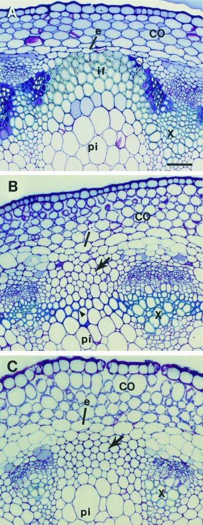
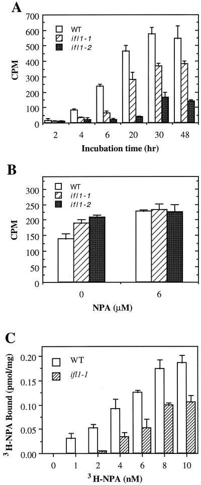
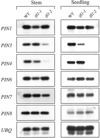
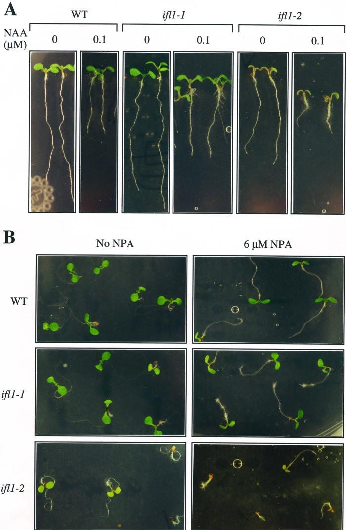
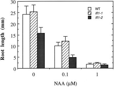
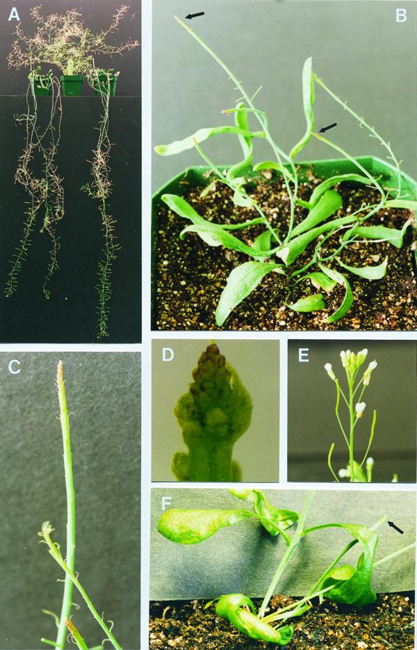
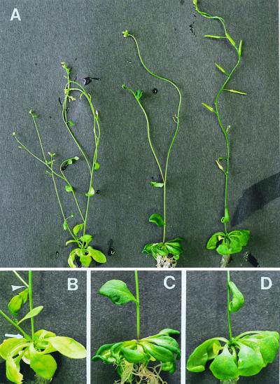
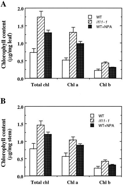
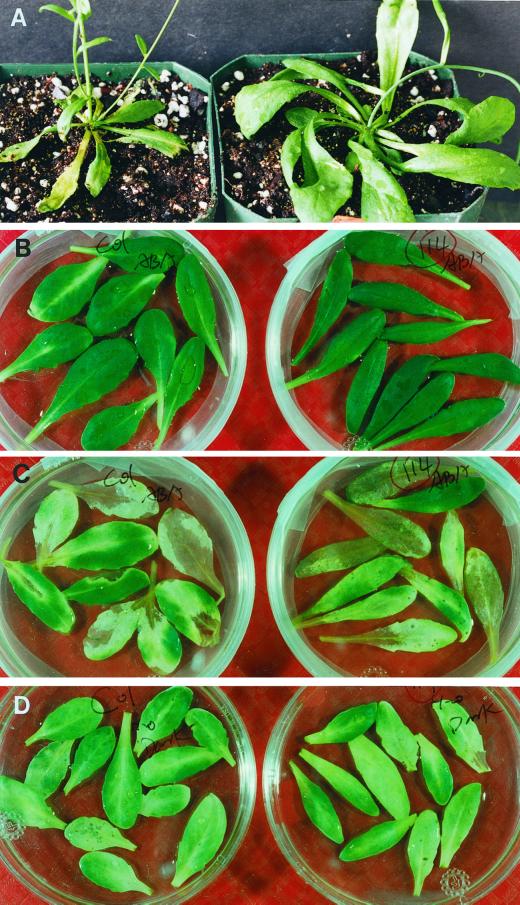
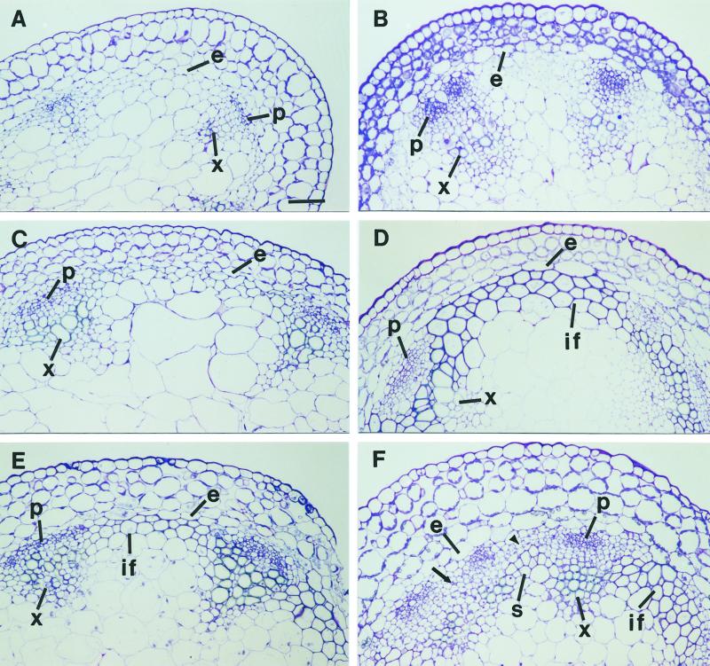
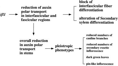
The INTERFASCICULAR FIBERLESS/REVOLUTA (IFL1/REV) gene is essential for the normal differentiation of interfascicular fibers and secondary xylem in the inflorescence stems of Arabidopsis. It has been proposed that IFL1/REV influences auxin polar flow or the transduction of auxin signal, which is required for fiber and vascular differentiation. Assay of auxin polar transport showed that the ifl1 mutations dramatically reduced auxin polar flow along the inflorescence stems and in the hypocotyls. The null mutant allele ifl1-2 was accompanied by a significant decrease in the expression level of two putative auxin efflux carriers. The ifl1 mutants remained sensitive to auxin and an auxin transport inhibitor. The ifl1-2 mutant exhibited visible phenotypes associated with defects in auxin polar transport such as pin-like inflorescence, reduced numbers of cauline branches, reduced numbers of secondary rosette inflorescence, and dark green leaves with delayed senescence. The visible phenotypes displayed by the ifl1 mutants could be mimicked by treatment of wild-type plants with an auxin polar transport inhibitor. In addition, the auxin polar transport inhibitor altered the normal differentiation of interfascicular fibers in the inflorescence stems of wild-type Arabidopsis. Taken together, these results suggest a correlation between the reduced auxin polar transport and the alteration of cell differentiation and morphology in the ifl1 mutants.


Figures

Figure 1
Figure 1
Disruption of interfascicular fiber differentiation in the inflorescence stems of ifl1. Sections were taken from the middle portions of inflorescence stems of 6-week-old plants and were stained with toluidine blue for anatomy. A, Section from the wild type showing the presence of four layers of interfascicular fibers located next to the endodermis. B, Section from ifl1-1 showing the interfascicular cells (arrow) next to the endodermis, which were normally destined to be fibers remained parenchymatous. However, ectopic sclerification occurred in some interfascicular cells (arrowhead), which were normally not destined to be sclerified. C, Section from ifl1-2 showing lack of any sclerified cells in the interfascicular region. co, Cortex; e, endodermis; if, interfascicular fiber; pi, pith; x, xylem. Bar (A) = 5 μm (A–C).
Figure 2
Figure 2
Auxin polar transport and NPA-binding activity in the wild type (WT) and ifl1. A, Time course of auxin polar transport activity in the inflorescence stems of wild type and ifl1. ifl1-1 and ifl1-2 mutants showed a dramatic reduction in the auxin polar transport activity compared with the wild type. Data are the mean values ± se of 15 plants. B, Auxin efflux in the hypocotyls of the wild type and ifl1. Auxin efflux rate in hypocotyl cells was measured by the retention of the amount of [3H] indole-3-acetic acid (IAA) in the presence or absence of NPA. The ifl1 mutants showed more retention of IAA compared with the wild type. Data are the mean values ± se of four replicates. C, 3H-NPA binding to plasma membranes prepared from the wild-type and ifl1-1 stems. Plasma membranes isolated from stems were incubated with various concentrations of 3H-NPA. Background binding activity was determined by addition of unlabeled NPA. The amount of 3H-NPA bound shown in the figure is the specific binding activity that was calculated by subtracting the background binding activity from total binding activity. ifl1-1 had a significant decrease in the NPA-binding activity compared with the wild type. Data are the mean values ± se of three assays.
Figure 3
Figure 3
Expression levels of PIN1 and other putative auxin efflux carrier genes in the wild type (WT) and ifl1 mutants. Total RNA isolated from stems or 2-week-old seedlings of the wild type and ifl1 mutants was used to analyze the expression levels of different genes by reverse transcriptase- (RT) PCR. The experiments were repeated three times and identical results were obtained. The expression level of ubiquitin (UBQ) gene was used as a control to confirm that equal amount of RNA was used for RT-PCR. The expression levels of PIN1, PIN6, PIN7, and PIN8 were similar in the stems and seedlings of the wild-type and ifl1 mutant, whereas the expression level of PIN3 and PIN4 was significantly reduced in ifl1-2 compared with the wild type.
Figure 4
Figure 4
Effects of NAA and NPA on the root growth of the wild type (WT) and ifl1 mutants. A, Display of the seedlings of the wild type and ifl1 mutants grown in agar medium containing no NAA or 1 μm NAA. NAA reduced root growth of wild-type and ifl1 mutants. B, Display of the seedlings of the wild-type and ifl1 mutants grown in agar medium containing no NPA or 6 μm NPA. NPA inhibited the downward growth and curling of roots of the wild-type and ifl1 mutants.
Figure 5
Figure 5
Inhibition of root growth of the wild-type (WT) and ifl1 mutants by NAA. Seeds were germinated on the agar medium containing 0, 0.1, or 1 μm NAA. Root length was measured 8 d after seedling growth. Each datum is the mean values ± se of 20 roots.
Figure 6
Figure 6
Morphological phenotypes displayed by ifl1-1 and ifl1-2. A, Prolonged inflorescence growth displayed by ifl1-1 (left and right) compared with the wild type (middle). B, Ifl1-2 showing pin-like inflorescence (arrows), dark green leaves, and reduced numbers of cauline branches. C, Close-up of ifl1-2 pin-like inflorescence showing lack of normal flowers and presence of thread-like structures. D, Close-up of ifl1-2 inflorescence apex showing lack of normal flower buds and presence of bulge-like structures. E, Wild-type inflorescence bearing normal flowers. F, pin1 mutant showing pin-like inflorescence (arrow), dark green leaves, and reduced numbers of cauline branches and secondary rosette inflorescence. These visible phenotypes resemble those seen in ifl1-2 (B and C).
Figure 7
Figure 7
The morphological phenotypes of ifl1 can be mimicked by growing wild-type plants in the presence of NPA. Plants were grown on solid Murashige and Skoog medium without or with the addition of 12 μm NPA for 7 weeks and were observed for their phenotypes. A, Morphology of wild type (left), wild type grown in the presence of NPA (middle), and ifl1-1. It was evident that ifl1-1 and NPA-treated wild type showed greener leaves and fewer cauline branches and secondary rosette inflorescence compared with the wild type. B, Close-up of the rosette of the wild type showing yellow leaves, cauline branch (arrowhead), and secondary rosette inflorescence (arrow). C, Close-up of the rosette of the NPA-treated wild type showing dark green and curling leaves and a lack of cauline branches and secondary rosette inflorescence. D, Close-up of the rosette of ifl1-1 showing dark green and curling leaves and a lack of cauline branches and secondary rosette inflorescence.
Figure 8
Figure 8
Chlorophyll content in the leaves and stems of the wild type (WT) and ifl1-1. Fresh leaves and stems were extracted in ethanol and the extracts were measured for chlorophyll content spectrophotometrically. Chlorophyll content was calculated based on fresh weight of leaves or stems. A, Measurement of chlorophyll content in leaves. B, Measurement of chlorophyll content in stems. ifl1-1 and wild type grown in the presence of NPA had much higher chlorophyll level than the wild type.
Figure 9
Figure 9
Senescence of wild-type and ifl1-1 leaves. Rosette leaves were detached from plants and incubated in liquid medium for test of senescence under darkness or treatment of ABA. At left is the wild type and at right is ifl1-1. A, Natural senescence of rosette leaves of 7-week-old plants. Although wild-type rosette leaves became yellow and dying (left), leaves of ifl1-1 (right) remained dark-green and alive. B, Freshly detached leaves displaying dark green color. C, Promotion of leaf senescence by treatment of ABA. It was evident that leaves of wild type and ifl1-1 were yellowing after treatment with ABA for 3 d. Leaves without ABA treatment remained green after 3 d of incubation (data not shown). D, Promotion of leaf senescence by darkness. It was obvious that leaves of wild type and ifl1-1 were yellowing after darkness treatment for 7 d.
Figure 10
Figure 10
Alteration of interfascicular fiber differentiation by treatment with the auxin polar transport inhibitor NPA. Plants were grown on the Murashige and Skoog medium with or without NPA. Inflorescence stems of 7-week-old plants were divided into three equal segments and the middle parts of stem segments were sectioned and stained with toluidine blue for anatomy. A and B, Sections from the top segments from plants grown on the medium without (A) or with (B) NPA. No secondary wall thickening was seen in the interfascicular cells. C and D, Sections from the middle segments from plants grown on the medium without (C) or with (D) NPA. Although interfascicular cells in plants treated with NPA (D) showed secondary wall thickening, those in plants without NPA treatment (C) did not. E and F, Sections from the basal segments from plants grown on the medium without (E) or with (F) NPA. Interfascicular fibers were evident in plants without NPA treatment (E), whereas some interfascicular regions in plants treated with NPA (F) were devoid of fibers (arrow and arrowhead) or had ectopic sclerification of interfascicular cells. e, Endodermis; if, interfascicular fiber; p, phloem; s, sclerenchyma; x, xylem. Bar (A) = 5 μm (A–F).
Figure 11
Figure 11
A tentative explanation of the phenotypes displayed in ifl1. The finding that the ifl1 mutations dramatically reduce auxin polar transport in the inflorescence stems led us to propose that some of the observed phenotypes were likely caused by an alteration in auxin polar flow along the stems. This hypothesis was further supported by the pharmacological studies that show that most of the ifl1 phenotypes could be mimicked by the treatment of wild-type plants with NPA.

References

    1. Aloni R. Polarity of induction and pattern of primary phloem fiber differentiation in Coleus. Am J Bot. 1976;63:877–889.
    1. Aloni R. Source of induction and sites of primary phloem fiber differentiation in Coleus blumei. Ann Bot. 1978;42:1261–1269.
    1. Aloni R. Role of auxin and gibberellin in differentiation of primary phloem fibers. Plant Physiol. 1979;63:609–614. - PMC - PubMed
    1. Aloni R. Role of cytokinin in differentiation of secondary xylem fibers. Plant Physiol. 1982;70:1631–1633. - PMC - PubMed
    1. Aloni R. Differentiation of vascular tissues. Annu Rev Plant Physiol. 1987;38:179–204.

Publication types