. 2001 Jun;126(2):601-12.
doi: 10.1104/pp.126.2.601.

The mitochondrial isovaleryl-coenzyme a dehydrogenase of arabidopsis oxidizes intermediates of leucine and valine catabolism

Affiliations

The mitochondrial isovaleryl-coenzyme a dehydrogenase of arabidopsis oxidizes intermediates of leucine and valine catabolism

K Däschner et al. Plant Physiol. 2001 Jun.

Abstract

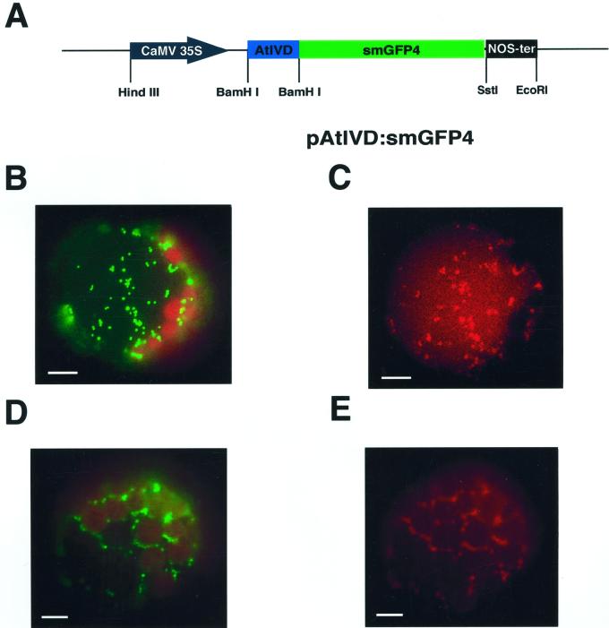
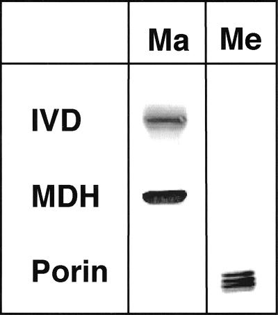
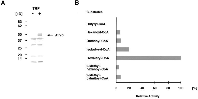
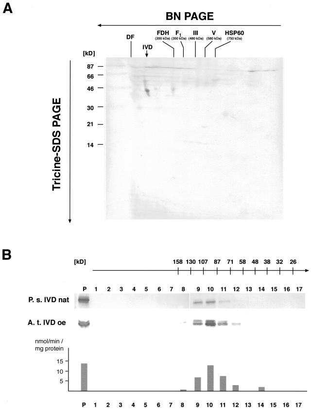
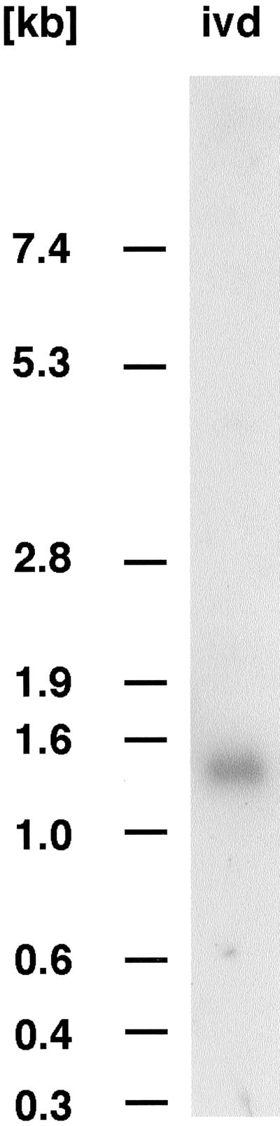
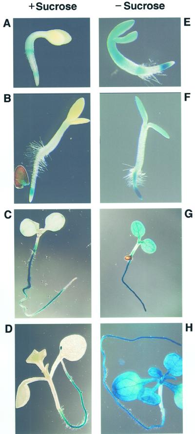
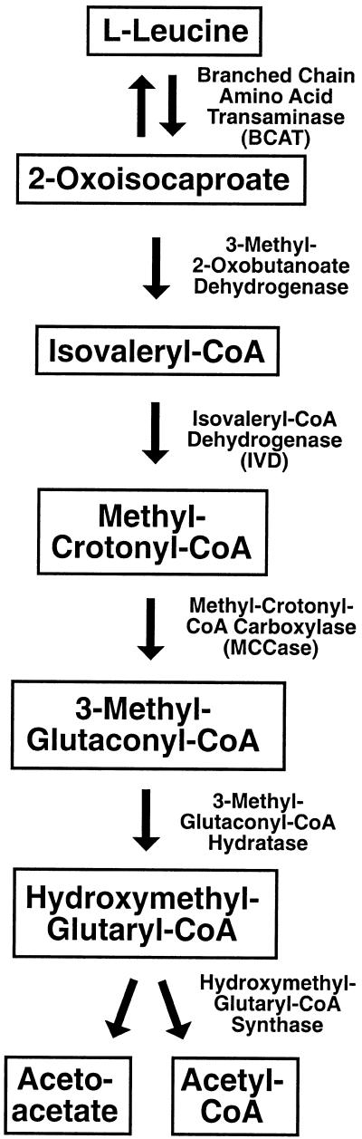
We recently identified a cDNA encoding a putative isovaleryl-coenzyme A (CoA) dehydrogenase in Arabidopsis (AtIVD). In animals, this homotetrameric enzyme is located in mitochondria and catalyzes the conversion of isovaleryl-CoA to 3-methylcrotonyl-CoA as an intermediate step in the leucine (Leu) catabolic pathway. Expression of AtIVD:smGFP4 fusion proteins in tobacco (Nicotiana tabacum) protoplasts and biochemical studies now demonstrate the in vivo import of the plant isovaleryl-CoA dehydrogenase (IVD) into mitochondria and the enzyme in the matrix of these organelles. Two-dimensional separation of mitochondrial proteins by blue native and SDS-PAGE and size determination of the native and overexpressed proteins suggest homodimers to be the dominant form of the plant IVD. Northern-blot hybridization and studies in transgenic Arabidopsis plants expressing Ativd promoter:gus constructs reveal strong expression of this gene in seedlings and young plants grown in the absence of sucrose, whereas promoter activity in almost all tissues is strongly inhibited by exogeneously added sucrose. Substrate specificity tests with AtIVD expressed in Escherichia coli indicate a strong preference toward isovaleryl-CoA but surprisingly also show considerable activity with isobutyryl-CoA. This strongly indicates a commitment of the enzyme in Leu catabolism, but the activity observed with isobutyryl-CoA also suggests a parallel involvement of the enzyme in the dehydrogenation of intermediates of the valine degradation pathway. Such a dual activity has not been observed with the animal IVD and may suggest a novel connection of the Leu and valine catabolism in plants.


Figures

Figure 1
Figure 1
An AtIVD:smGFP4 fusion protein is imported into mitochondria of tobacco protoplasts. A, Construct carrying the gene encoding AtIVD:smGFP4 fusion protein. The N-terminal part of the Ativd cDNA encoding the first 50 amino acids (AtIVD, blue box) is cloned upstream of the smGFP4 reading frame (smGFP4, green box). Expression of the resulting fusion protein is controlled by the cauliflower mosaic virus 35S promoter (black arrow) and the NOS terminator (black box). Restriction sites are indicated for HindIII, BamHI, SstI, and EcoRI. B through E, Tobacco protoplasts expressing the AtIVD:smGFP4 fusion protein (B and C) and CPN-60:mGFP5 (D and E). Images B and D were taken with flourescein isothiocyanat (BP 450–490/LP515) and C and E were analyzed with MitoTracker (HQ545/30/HQ 610/75) filter sets, respectively. The green fluorescent protein (GFP) is observed in particles with sizes of about 1 μm. The colocalization of the GFP and MitoTracker Red fluorescence confirms the localization of the fusion proteins in mitochondria. The bars given in the individual frames correspond to 10 μm.
Figure 2
Figure 2
The AtIVD is located in the mitochondrial matrix. Mitochondria isolated from etiolated pea seedlings were fractionated into membrane (Me) and matrix (Ma) fractions. The quality of the fractionation was confirmed by immunodetection with antibodies against the mitochondrial matrix protein malate dehydrogenase from watermelon (Citrullus lanatus; MDH) and the outer membrane protein porin of potato (Solanum tuberosum; Porin). Immunodetection using an antibody against the human mitochondrial IVD decorates a protein of 43 kD only in the matrix fraction confirming the presence of the IVD in the mitochondrial matrix space.
Figure 3
Figure 3
Substrate specificity of the overexpressed AtIVD. A, Western-blot analysis of the overexpressed AtIVD with an IVD-specific antibody against the homologous protein from human. A clear signal corresponding to the expected size of about 43 kD (highlighted by an arrow and designated AtIVD) is specifically detected only after induction (lane +) with Trp (TRP) but is not observed in noninduced E. coli cells (lane −). B, Relative activities (given in %) measured with different acyl-CoA substrates at 50-μm final concentration in a typical enzyme test. Highest activity as found with isovaleryl-CoA is arbitrarily set at 100%. Values correspond to those given in Table I, first column.
Figure 4
Figure 4
The plant IVD is a homodimer. A, Two-dimensional separation of Arabidopsis mitochondrial proteins. Mitochondrial proteins obtained from an Arabidopsis tissue culture were separated under native conditions in the presence of Coomassie Blue in the first dimension (BN-PAGE). A lane containing the size-separated protein complexes was cut out and transferred to Tricine-SDS-PAGE in the second dimension. Proteins were subsequently transferred to polyvinylidene difluoride membranes and investigated by immunostaining. A spot corresponding to the IVD polypeptide with 43 kD is detected with the IVD-specific antibody (indicated by a vertical arrow). The migration behavior of the protein in the first dimension indicates a native size of about 60 to 100 kD, suggesting a mono- or dimeric native structure of the protein. Masses of native respiratory chain complexes in the first dimension are given for fumarate dehydrogenase (FDH), the F1 part of complex V (F1), complexes III (III) and V (V), and the CPN-60 complex. The position of the dye front (DF) is also indicated. Molecular masses of marker proteins co-electrophoresed in the second dimension are given in kD on the left-hand side. B, Gel filtration chromatography of pea mitochondrial proteins and E. coli proteins containing overexpressed AtIVD. The apparent sizes (given in kD above the numbering of the fractions) expected in the individual fractions are deduced from the calibration of the column with marker proteins. Proteins of the individual fractions are investigated by western-blot/immunostaining analysis with an IVD-specific antibody. The native pea IVD (P.s. IVD nat, upper) as well as overexpressed AtIVD (A. t. IVD oe, lower) are detected in the protein fraction prior to separation (P) and in fractions corresponding to proteins with molecular masses between 130 and 71 kD with the majority of the protein eluting in fraction 10 corresponding to proteins between 107 and 87 kD. This is about twice the molecular mass of the plant IVD corroborating the dimeric form of the plant IVD. Enzyme activity (given in nmol min−1 mg−1 protein) of eluted fractions containing overexpressed AtIVD was measured with isovaleryl-CoA. Levels of activity correspond to the amounts of detected IVD protein indicating that the protein remained in its active state during the gel filtration analysis.
Figure 5
Figure 5
Northern-blot analysis of total Arabidopsis RNAs from aboveground tissues. The hybridization with a probe corresponding to the complete Ativd cDNA detects a single RNA species of about 1.4 to 1.5 kb. Sizes of co-electrophoresed RNA marker molecules are given in kb.
Figure 6
Figure 6
Ativd promoter activity is significantly inhibited by Suc. A through H, GUS staining of Arabidopsis seedlings grown in the presence (A–D) or absence (E–H) of 0.5% (w/v) Suc. In early developmental stages (A and B), expression observed in seedlings grown in the absence of Suc is rather low. In later stages, however, clear differences in Ativd promoter activity become apparent between seedlings grown in the absence (G and H) or presence (C and D) of Suc. No influence of Suc is observed in root tissues. Seedlings correspond to stages 2 (A and E), 3 (B and F), 7 (C and G), and 15 (D and H) d after sowing.
Figure 7
Figure 7
Scheme of the catabolic pathway of Leu. Intermediates are given in boxes. All enzymatic activities (given at the right-hand sides of the vertical arrows) have been crudely detected in soybean (Anderson et al., 1998) and are well characterized in animals.

References

    1. Alban C, Baldet P, Axiotis S, Douce R. Purification and characterization of 3-methylcrotonyl-coenzyme A carboxylase from higher plant mitochondria. Plant Physiol. 1993;102:957–965. - PMC - PubMed
    1. Anderson MD, Che P, Song J, Nikolau BJ, Wurtele ES. 3-Methylcrotonyl-coenzyme A carboxylase is a component of the mitochondrial leucine catabolic pathway in plants. Plant Physiol. 1998;118:1127–1138. - PMC - PubMed
    1. Aubert S, Alban C, Bligny R, Douce R. Induction of β-methylcrotonyl-coenzyme A carboxylase in higher plant cells during carbohydrate starvation: evidence for a role of MCCase in leucine catabolism. FEBS Lett. 1996;383:175–180. - PubMed
    1. Bechtold N, Ellis J, Peletier G. In planta Agrobacterium mediated gene transfer by infiltration of adult Arabidopsis thaliana plants. C R Acad Sci. 1993;316:1194–1199.
    1. Binder S, Brennicke A. Transcription initiation sites in mitochondria of Oenothera berteriana. J Biol Chem. 1993;268:7849–7855. - PubMed

Publication types

MeSH terms