. 2001 Jun;126(2):643-55.
doi: 10.1104/pp.126.2.643.

The arabidopsis ATHB-8 HD-zip protein acts as a differentiation-promoting transcription factor of the vascular meristems

Affiliations

The arabidopsis ATHB-8 HD-zip protein acts as a differentiation-promoting transcription factor of the vascular meristems

S Baima et al. Plant Physiol. 2001 Jun.

Abstract

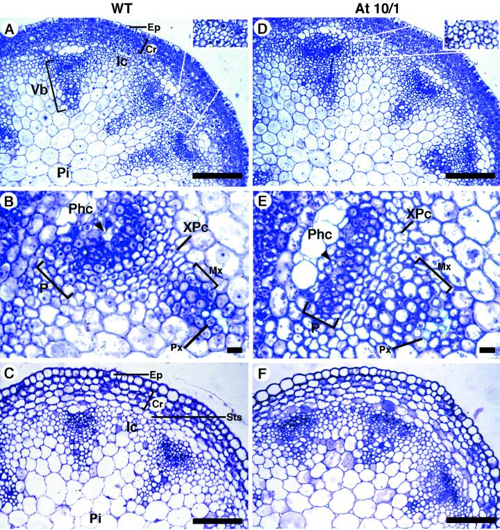
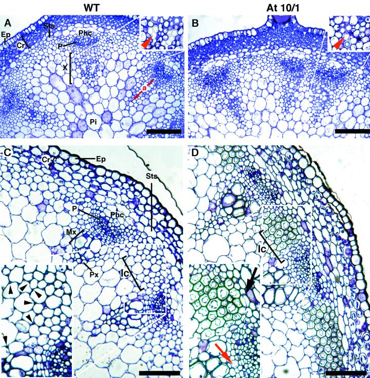
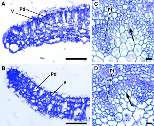
ATHB-8, -9, -14, -15, and IFL1/REV are members of a small homeodomain-leucine zipper family whose genes are characterized by expression in the vascular tissue. ATHB-8, a gene positively regulated by auxin (Baima et al., 1995), is considered an early marker of the procambial cells and of the cambium during vascular regeneration after wounding. Here, we demonstrate that although the formation of the vascular system is not affected in athb8 mutants, ectopic expression of ATHB-8 in Arabidopsis plants increased the production of xylem tissue. In particular, a careful anatomical analysis of the transgenic plants indicated that the overexpression of ATHB-8 promotes vascular cell differentiation. First, the procambial cells differentiated precociously into primary xylem. In addition, interfascicular cells also differentiated precociously into fibers. Finally, the transition to secondary growth, mainly producing xylem, was anticipated in transgenic inflorescence stems compared with controls. The stimulation of primary and secondary vascular cell differentiation resulted in complex modifications of the growth and development of the ATHB-8 transgenic plants. Taken together, these results are consistent with the hypothesis that ATHB-8 is a positive regulator of proliferation and differentiation, and participates in a positive feedback loop in which auxin signaling induces the expression of ATHB-8, which in turn positively modulates the activity of procambial and cambial cells to differentiate.


Figures

Figure 1
Figure 1
Structure of the ATHB-8 gene, En-1 transposon insertion sites, and ATHB-8 expression in athb-8 mutants. A, Schematic drawing of the ATHB-8 gene. The gene has 17 introns, with the first intron located in the 5′ untranslated region just before the start codon. Introns are shown as lines and exons as boxes. White boxes, 5′ and 3′ untranslated region; sketch boxes, HD-Zip domain; gray boxes, START domain. The positions of the two independent En-1 insertions are indicated by white arrowheads. B, Northern-blot analysis of ATHB-8 expression. A DNA fragment corresponding to the 3′ region of the ATHB-8 cDNA (a 272-bp EcoRI DNA fragment; Baima et al., 1995) was used to probe 10 μg of total RNA isolated from 2-week-old Arabidopsis plants. The same blot was probed with a cDNA for the β-subunit of N. plumbaginifolia mitochondrial ATPase (βATPase).
Figure 2
Figure 2
Morphology of soil-grown ATHB-8 transgenic plants. A through C, Side view of 4-week-old wild-type (A), At5/2 (B), and At10/1 (C) plants. D through F, Root architecture of 6-week-old wild-type (D), At5/2 (E), and At10/1 (F) plants. Scale bar = 3 mm.
Figure 3
Figure 3
Histological analysis of the secondary vascular structure in the inflorescence stem of ATHB-8 transgenic plants. A, Transverse section of the basal end of the stem showing the onset of the activity of the interfascicular (arrows) and fascicular (arrowheads) cambium in 5-week-old wild-type plants. B, Schematic diagram showing the organization of the Arabidopsis vascular system in stem undergoing secondary growth: phloem (green), fascicular xylem (purple), interfascicular xylem (dark blue), interfascicular fiber cells (light blue); the arrows indicate the radius of the: (a) fascicular xylem, (b) middle part of the interfascicular arc, and (c) interfascicular arc flanking the bundle. The average measurements are indicated in Table I. C–E, Transverse sections of the basal end of stem showing the secondary structure of the vascular system in 6-week-old stem. C, Wild type. D, At5/2. E, At10/1. F through I, Transverse sections of the basal part of the stem were taken from 6-week-old (F and G) or 8-week-old (H and I) plants showing other secondary growth events peculiar to ATHB-8 transgenic plants. G, Significant production of phloem fiber sclereids (arrowhead); I, lignification of a wide part of the pith was observed in the stem of At10/1 (G and I) and not in the wild type (F and H). Sections were stained with toluidine blue (A–E) and carmin-iodine mixture (F and G). H and I, Autofluorescence of lignified cell walls. Scale bar: 100 μm (A–E, H, and I) and 20 μm (F and G). Ep, Epidermis; Cr, cortex; Ic, interfascicular cells; Mx, metaxylem; P, phloem; Pi, pith; Px, protoxylem; Sts, starch sheath; Sx, secondary xylem.
Figure 4
Figure 4
Histological analysis of the secondary vascular structure in the root of ATHB-8 plants. A, Schematic diagram showing secondary growth in the Arabidopsis root: xylem (dark purple), cambium and phloem (green), periderm (yellow); the arrows indicate the radius of the (d) vascular region, (e) periderm, and (f) entire root. B through D, Transverse sections showing the degree of secondary vascular growth in wild-type (B), At5/2 (C), and At10/1 (D) root. The transverse sections were taken from the region immediately below the hypocotyl-root junction of 6-week-old plants grown on soil. Sections were stained with toluidine blue. Scale bar: 100 μm.
Figure 5
Figure 5
Histological analysis of the primary vascular structure in 1-cm-long inflorescence stem of ATHB-8 transgenic plants. A through C, Wild type. D through F, At10/1. A and D, Transverse sections showing representative bundles of the apical part of 1-cm-long stem (eustele); the insets show magnifications of the interfascicular region indicating a lower meristematic activity of the transgenic plant compared with the control. B and E, Magnification of typical vascular bundles. Note the absence of radially aligned immature xylem cells and the flattened phloem in the transgenic primary bundle. The arrowheads show protophloem cells. C and F, Transverse sections showing representative bundles of the basal part of 1-cm-long stem. Sections were stained with toluidine blue. Scale bar: 100 μm (A, C, D, and F) and 10 μm (B and E). Ep, Epidermis; Cr, cortex; Ic, interfascicular cells; Mx, metaxylem; P, phloem; Phc, phloem cap cells; Pi, pith; Px, protoxylem; Sts, starch sheath; Vb, vascular bundle; XPc, xylary procambium.
Figure 6
Figure 6
Histological analysis of the vascular structure in 5-cm-long inflorescence stem of ATHB-8 transgenic plants. A and C, Wild type. B and D, At10/1. A and B, Transverse sections showing representative bundles of the apical part of 5-cm-long stem (eustele). The red arrow (a) indicates the radius of the primary xylem considered for statistical analysis. A magnification of the protoxylem cells (indicated by red arrowhead in the insets) shows a higher secondary wall deposition in the transgenic plant compared with the control. C and D, Transverse sections showing representative bundles of the basal end of 5-cm-long stem. Note the deposition of secondary wall thickenings (black arrowheads) at the angles of the interfascicular cells in C, compared with the lignified interfascicular cells in D. The onset of the interfascicular cambium (black arrow) and a secondary xylem cell (red arrow) are also shown in D. Sections were stained with toluidine blue. Scale bar: 100 μm (A–D). Ep, Epidermis; Cr, cortex; Ic, interfascicular cells; Mx, metaxylem; P, phloem; Phc, phloem cap cells; Pi, pith; Px, protoxylem; Sts, starch sheath; Sx, secondary xylem; X, xylem.
Figure 7
Figure 7
Histological analysis of the primary vascular structure in leaf and pedicel of ATHB-8 transgenic plants. A and C, Wild type. B and D, At10/1. A and B, Transverse sections showing a lateral portion of the lamina. Sections were taken at the middle of the seventh leaf of 3-week-old plants. Note the presence of meristematic tissue at the margins of the transgenic leaf in B. C and D, Transverse sections showing differentiating procambial traces in flower pedicels. In the interfascicular region (arrow) the meristematic component is higher in the transgenic plants than in the control. Sections were stained with toluidine blue. Scale bar: 50 μm (A and B and 10 μm (C and D). Pd, Palisade cells; Pt, procambial traces; V, vein.

References

    1. Aloni R. Differentiation of vascular tissues. Annu Rev Plant Physiol. 1987;38:179–204.
    1. Altamura MM, Possenti M, Matteucci A, Baima S, Ruberti I, Morelli G (2001) The development of the vascular system in the Arabidopsis floral stem. New Phytol (in press) - PMC - PubMed
    1. Aoyama T, Dong C-H, Wu Y, Carabelli M, Sessa G, Ruberti I, Morelli G, Chua N-H. Ectopic expression of the Arabidopsis transcriptional activator ATHB-1 alters leaf cell fate in tobacco. Plant Cell. 1995;7:1773–1785. - PMC - PubMed
    1. Baima S, Nobili F, Sessa G, Lucchetti S, Ruberti I, Morelli G. The expression of the ATHB-8 homeobox gene is restricted to provascular cells in Arabidopsis thaliana. Development. 1995;121:4171–4182. - PubMed
    1. Baima S, Tomassi M, Matteucci A, Altamura MM, Ruberti I, Morelli G. Role of the ATHB-8 gene in xylem formation. In: Savidge R, Barnett J, Napier R, editors. Cambium: The Biology of Wood Formation. Oxford: βIOS Scientific Publishers LTD; 2000. pp. 445–455.

Publication types

MeSH terms