. 2001 Jun;126(2):717-30.
doi: 10.1104/pp.126.2.717.

The Arabidopsis embryo mutant schlepperless has a defect in the chaperonin-60alpha gene

Affiliations

The Arabidopsis embryo mutant schlepperless has a defect in the chaperonin-60alpha gene

N R Apuya et al. Plant Physiol. 2001 Jun.

Abstract

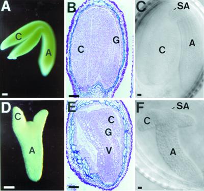
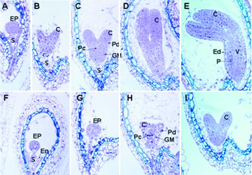
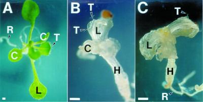
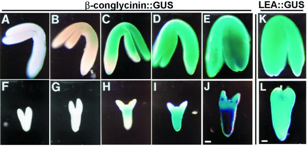
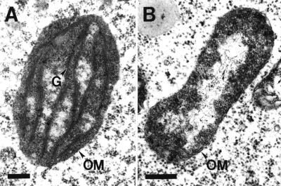
We identified a T-DNA-generated mutation in the chaperonin-60alpha gene of Arabidopsis that produces a defect in embryo development. The mutation, termed schlepperless (slp), causes retardation of embryo development before the heart stage, even though embryo morphology remains normal. Beyond the heart stage, the slp mutation results in defective embryos with highly reduced cotyledons. slp embryos exhibit a normal apical-basal pattern and radial tissue organization, but they are morphologically retarded. Even though slp embryos are competent to transcribe two late-maturation gene markers, this competence is acquired more slowly as compared with wild-type embryos. slp embryos also exhibit a defect in plastid development-they remain white during maturation in planta and in culture. Hence, the overall developmental phenotype of the slp mutant reflects a lesion in the chloroplast that affects embryo development. The slp phenotype highlights the importance of the chaperonin-60alpha protein for chloroplast development and subsequently for the proper development of the plant embryo and seedling.


Figures

Figure 1
Figure 1
Morphology of schlepperless embryos. Mature embryos from WT (A, B, and C) and schlepperless (D, E, and F). Whole mount photographs of embryos dissected out of the same mature silique (A and D). Sections of embryos from mature silique embedded in LR White plastic resin (B and E). Nomarski photographs of embryos taken from the same mature silique (C and F). C, Cotyledon; Ep, epidermis; G, ground tissue; H, hypocotyl; SA, shoot apical meristem; V, vascular tissue. Bars = 50 μm.
Figure 2
Figure 2
Developmental analysis of schlepperless embryos. A developmental series of sections for Arabidopsis WT (A–E) and schlepperless (F–I) embryos embedded in LR White plastic resin. The mutant embryos were from the same silique from which the corresponding WT embryos were taken. A, Axis; C, cotyledon; EP, embryo proper; Ep, epidermis; En, endosperm; GM, ground meristem; P, storage parenchyma; Pc, procambium; Pd, protoderm; S, suspensor; V, vascular tissue.
Figure 3
Figure 3
Germination of WT and schlepperless seeds in vitro. A, WT seedling after 12 d in culture; B and C, schlepperless seedling after 42 (B) and after 70 (C) d in culture. The mutant seedling shown in C was the same seedling shown in B. C, Cotyledon; H, hypocotyl; L, leaf; R, root; T, trichome. Bars = 50 μm.
Figure 4
Figure 4
schlepperless embryo is competent to transcribe late-maturation genes. Expression pattern of β-conglycinin::GUS transgene in WT (A–E) and schlepperless (F–J) embryos at different developmental stages. Expression pattern of LEA::GUS transgene in mature WT (K) and schlepperless (L) embryos. Corresponding WT and mutant embryos were taken from the same silique. Blue staining indicates activity of GUS. Bars = 50 μm.
Figure 5
Figure 5
The Chaperonin-60α gene is interrupted by T-DNA in slp mutant. A, Diagrammatic representation of the WT chaperonin-60α locus and the concatemer of T-DNAs inserted in slp line (not drawn to scale). Exons are shown as unshaded boxes superimposed on the heavy line. The 11-kb WT genomic clone and the 6.6-kb EcoRI sub-clone are indicated by thin lines. The insertion point of the T-DNA concatemer is indicated by two lines below the seventh exon. Left border (LB) and right border (RB) regions are indicated in the adjacent rectangles that represent the T-DNA concatemer (see Errampalli et al., 1991, for the T-DNA map). Numbers in shaded rounded rectangles indicate the expected size of some polymorphic fragments as shown in the genomic blots (B and C). Not all of the restriction sites within the T-DNA are shown. E, EcoRI; H, HindIII, S, SalI. B, DNA gel-blot analysis of WT and heterozygous (HZ) individuals using right border (first) or a BamHI fragment representing a portion of the RB-plant-flanking region (second). Relevant fragments are indicated by fragment size corresponding to the map in (A). C, Genomic DNA-blot analysis of randomly selected F2 plants used in the genetic analysis presented in Table I. The probe used was the partial pC31 cDNA clone. Genomic DNAs digested with EcoRI were electrophoresed in 1% (w/v) agarose gel, whereas those digested with NotI were electrophoresed in 0.5% (w/v) agarose gel. Samples from genotypically WT (WT) and heterozygous (HZ) individuals were included as controls.
Figure 6
Figure 6
Alignment of the Arabidopsis chaperonin-60α protein with other related proteins. The Arabidopsis chaperonin-60α protein was aligned with chaperonin-60α protein from B. napus (95% similarity), Arabidopsis chaperonin-60β protein (70% similarity), the Arabidopsis mitochondrial chaperonin-60 (90% similarity), and GroEL protein from Escherichia coli (70% similarity) using the AlignX program of VectorNTI software. Amino acids that are identical in all five proteins are presented in yellow blocks with red letters. Conservative amino acids are presented in gray blocks with black letters. Sources of these proteins are cited in the text. The genomic sequence of the Arabidopsis Chaperonin-60α has the GenBank accession no. U49357.
Figure 7
Figure 7
RNA-blot analysis of chaperonin-60α. Each lane contains 0.5 μg of poly(A+) mRNA from each organ. The RNA blot was hybridized with the 6.6-kb EcoRI fragment from λ101 genomic clone (see Fig. 5A).
Figure 8
Figure 8
Transmission electron microscopy analysis of plastids. Chloroplast (A) from a WT embryo and undeveloped plastid (B) from slp embryo. G, Grana; OM, outer membrane. Bars = 0.15 μm.

Similar articles

Cited by

References

    1. Ausubel FM, Brent R, Kingston RE, Moore DD, Seidman JG, Smith JA, Struhl K, editors. Current Protocol in Molecular Biology. New York: Greene Publishing and Wiley-Interscience; 1992.
    1. Behringer FJ, Medford JI. A plasmid rescue technique for the recovery of plant DNA disrupted by T-DNA insertion. Plant Mol Biol Rep. 1992;10:190–198.
    1. Berleth T, Jürgens G. The role of monopteros in organizing the basal body region of the Arabidopsis embryo. Development. 1993;118:575–587.
    1. Castle LA, Errampalli D, Aterton TL, Franzmann LH, Yoon ES, Meinke DW. Genetic and molecular characterization of embryonic mutants identified following seed transformation in Arabidopsis. Mol Gen Genet. 1993;241:504–514. - PubMed
    1. Clark JK, Sheridan WF. Isolation and characterization of 51 embryo-specific mutations in maize. Plant Cell. 1991;3:935–951. - PMC - PubMed