. 2001 Jun;126(2):801-10.
doi: 10.1104/pp.126.2.801.

Molecular characterization of At5PTase1, an inositol phosphatase capable of terminating inositol trisphosphate signaling

Affiliations

Molecular characterization of At5PTase1, an inositol phosphatase capable of terminating inositol trisphosphate signaling

S E Berdy et al. Plant Physiol. 2001 Jun.

Abstract

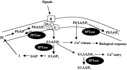
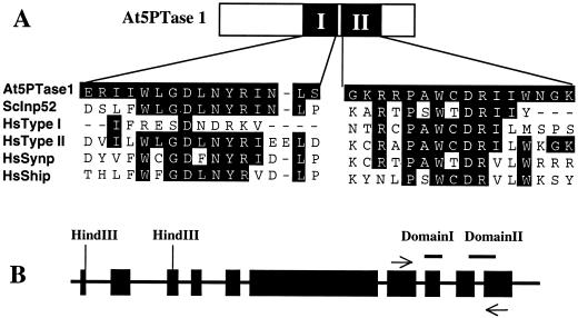
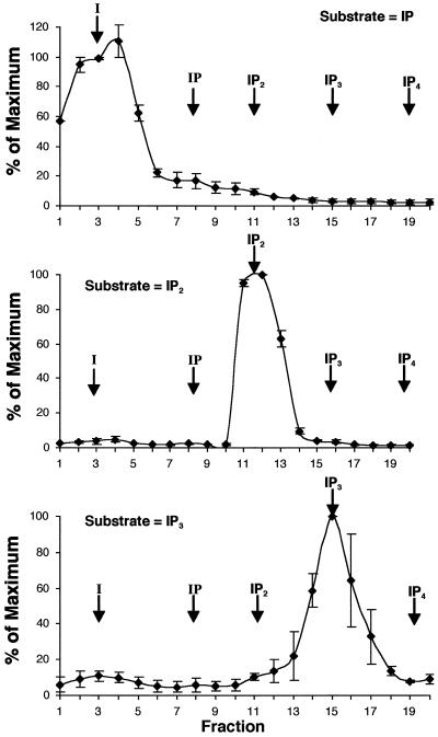
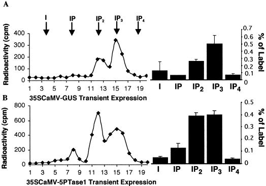
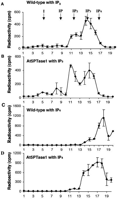
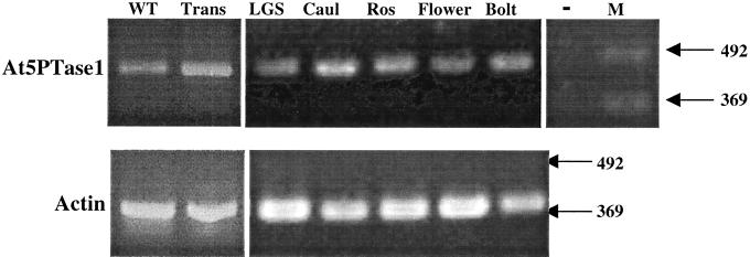
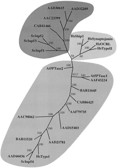
The inositol triphosphate (IP(3))-signaling pathway has been associated with several developmental and physiological processes in plants, but we currently know little about the regulation of this pathway. Inositol 5' phosphatases (5PTases) are enzymes that remove a 5' phosphate from several potential second messengers, including IP(3). In catalyzing the removal of a 5' phosphate from second messenger substrates, 5PTases can act to terminate signal transduction events. We describe the molecular analysis of At5PTase1, a 5PTase gene from Arabidopsis. When expressed transiently in Arabidopsis leaf tissue or ectopically in transgenic plants, At5PTase1 allowed for the increased hydrolysis of I(1,4,5)P(3) and I(1,3,4,5)P(4) substrates. At5PTase1 did not hydrolyze I(1)P, I(1,4)P(2), or PI(4,5)P(2) substrates. This substrate specificity was similar to that of the human Type I 5PTase. We identified 14 other potential At5PTase genes and constructed an unrooted phylogenetic tree containing putative Arabidopsis, human, and yeast 5PTase proteins. This analysis indicated that the Arabidopsis 5PTases were grouped in two separate branches of the tree. The multiplicity of At5PTases indicates that these enzymes may have different substrate specificities and play different roles in signal termination in Arabidopsis.


Figures

Figure 1
Figure 1
Signaling via IP3 and signal termination via 5PTases. Extracellular signals are perceived by putative receptors (R) that stimulate phospholipase C (PLC)/G protein complexes (G) to convert substrate PIP2 into the second messenger IP3. IP3 and related second messengers alter intracellular Ca2+ levels, thereby triggering downstream biological events. Termination of signaling can occur by hydrolysis of four different second messengers by 5PTase enzymes (black ovals).
Figure 2
Figure 2
Structure of At5PTase1 predicted protein sequence and alignment with known 5PTases. A, Schematic representation of At5PTase1 protein. The catalytic region consists of two conserved domains (I and II) found in all identified 5PTase proteins (Communi and Erneux, 1996; Communi et al., 1996; Jefferson and Majerus, 1996). B, Genomic structure of the At5PTase1-coding region. The genomic sequence and cDNA corresponding to At5PTase1 were compared using BLAST and Lasergene software. The black boxes correspond to the 10 identified exons. Exon sequences corresponding to domains I and II of the catalytic region are indicated by the bars on top; the location of PCR primers is noted by the arrows.
Figure 3
Figure 3
LeIMP-2 is a specific monophosphatase. Bacterial extracts expressing LeIMP2 were incubated with radiolabeled inositol phosphate substrates as described. Inositol and inositol phosphates were separated with SepPak columns using a triethylammonium buffer gradient. Twenty-one-milliliter fractions were collected and a portion was analyzed by scintillation counting. The mean value from three experiments was determined and normalized by comparison with the maximal value obtained. Arrows indicate the position of inositol phosphate peaks obtained when non-recombinant bacterial extracts were incubated with radiolabeled substrates.
Figure 4
Figure 4
Transient expression of At5PTase1 yields an active 5PTase. Arabidopsis leaf tissue was vacuum infiltrated with Agrobacterium tumefaciens containing a 35SCaMV-GUS construct (A) or a 35SCaMV-At5PTase1 construct (B) and tissue was extracted 24 h later. Extracts were incubated with H3-I(1,4,5)P3 substrate and the products separated over SepPak columns with a triethylammonium buffer gradient as described. Chromatograms are presented on the left. The arrows indicate the peak position of inositol and inositol phosphates as determined in Figure 3. The total radioactivity present in each peak was calculated for two separate experiments and is presented as mean and sd on the right.
Figure 5
Figure 5
Ectopic expression of At5PTase1 yields an active 5PTase. Protein extracts were isolated from Arabidopsis wild type (A and C) and transgenic plants expressing At5PTase1 under control of the 35SCaMV promoter (B and D) and were incubated with H3-I(1,4,5)P3 (A and B) or H3-I(1,3,4,5)P4 (C and D) substrate. Products were separated over SepPak columns with a triethylammonium buffer gradient as described. The total radioactivity present in each fraction was calculated for two separate experiments and is presented as mean and sd. The arrows indicate the peak position of inositol and inositol phosphates.
Figure 6
Figure 6
RT-PCR analysis of At5PTase1 expression. cDNA was synthesized from 5 μg of wild-type rosette leaves (WT), At5PTase1 transgenic leaves (Trans), light-grown seedling (LGS), cauline leaf (Caul), rosette leaf (Ros), flower, and bolt RNAs with Murine Moloney Leukemia Virus-RT and oligo dT primer. After dilution, cDNAs were amplified with Taq polymerase using At5PTase1 or actin primers for 30 cycles (94oC, 1 min, 56°C 1.5 min, and 72°C, 1 min) and were analyzed by gel electrophoresis. The At5PTase1 negative control (−) contained no RNA template. In addition, no products were amplified in reactions using each At5PTase 1 or actin primer alone. A 100-bp DNA ladder was used as a marker (M).
Figure 7
Figure 7
Phylogenetic analysis of 5PTase proteins. 5PTase protein sequences were aligned with ClustalW software. Alignments were analyzed with the ProtPars function of Phylip available by web interface on the Institute Pasteur Server (http://bioweb.pasteur.fr/seqanal/phylogeny/intro-uk.html) to generate an unrooted tree. Percentage bootstrap values of 500 replicates are given at each branch point. Branch lengths are to scale. Accession numbers are given for all except Type I 5PTase (CAA54676), Type II 5PTase (P32019), OCRL (AAB03839), Synaptojanin 1 (AAC51921), Ship 1 (AAB53573), Inp51(NP 012264), Inp52 (NP 014293), Inp53 (NP 014752), and Inp54 (NP 014576). At5PTase2 and CAB41466 were previously identified in the database as putative 5PTase genes. Accompanying information on size and substrate specificity can be found online in the supplemental data.

Similar articles

Cited by

References

    1. Berridge MJ. Inositol trisphosphate and calcium signaling. Nature. 1993;361:315–325. - PubMed
    1. Chandra S, Low PS. Measurement of Ca2+fluxes during elicitation of the oxidative burst in aequorin-transformed tobacco cells. J Biol Chem. 1997;272:28274–28280. - PubMed
    1. Clough SJ, Bent AF. Floral dip: a simplified method for Agrobacterium-mediated transformation of Arabidopsis thaliana. Plant J. 1998;16:735–743. - PubMed
    1. Communi D, Erneux C. Identification of an active site cysteine residue in human type I Ins(1,4,5) P35-phosphatase by chemical modification and site-directed mutagenesis. Biochem J. 1996;320:181–186. - PMC - PubMed
    1. Communi D, Lecocq R, Erneux C. Arginine 343 and 350 are two active residues involved in substrate binding by human Type I D-myo-inositol 1,4,5,-trisphosphate 5-phosphatase. J Biol Chem. 1996;271:11676–11683. - PubMed

Publication types