

Proteomic analysis of arabidopsis seed germination and priming
- PMID: 11402211
- PMCID: PMC111173
- DOI: 10.1104/pp.126.2.835
Proteomic analysis of arabidopsis seed germination and priming
Abstract
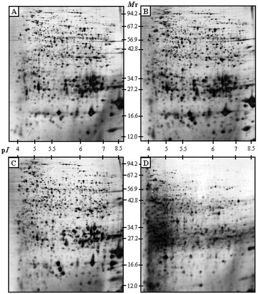
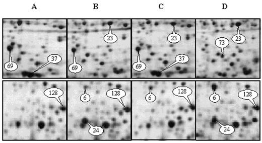
To better understand seed germination, a complex developmental process, we developed a proteome analysis of the model plant Arabidopsis for which complete genome sequence is now available. Among about 1,300 total seed proteins resolved in two-dimensional gels, changes in the abundance (up- and down-regulation) of 74 proteins were observed during germination sensu stricto (i.e. prior to radicle emergence) and the radicle protrusion step. This approach was also used to analyze protein changes occurring during industrial seed pretreatments such as priming that accelerate seed germination and improve seedling uniformity. Several proteins were identified by matrix-assisted laser-desorption ionization time of flight mass spectrometry. Some of them had previously been shown to play a role during germination and/or priming in several plant species, a finding that underlines the usefulness of using Arabidopsis as a model system for molecular analysis of seed quality. Furthermore, the present study, carried out at the protein level, validates previous results obtained at the level of gene expression (e.g. from quantitation of differentially expressed mRNAs or analyses of promoter/reporter constructs). Finally, this approach revealed new proteins associated with the different phases of seed germination and priming. Some of them are involved either in the imbibition process of the seeds (such as an actin isoform or a WD-40 repeat protein) or in the seed dehydration process (e.g. cytosolic glyceraldehyde-3-phosphate dehydrogenase). These facts highlight the power of proteomics to unravel specific features of complex developmental processes such as germination and to detect protein markers that can be used to characterize seed vigor of commercial seed lots and to develop and monitor priming treatments.
Figures
References
-
- Alban C, Job D, Douce R. Biotin metabolism in plants. Annu Rev Plant Physiol Plant Mol Biol. 2000;51:17–47. - PubMed
-
- Baldauf SL, Palmer JD. Evolutionary transfer of the chloroplast tufA gene to the nucleus. Nature. 1990;344:262–265. - PubMed
-
- Bewley JD, Black M. Seeds: Physiology of Development and Germination. New York: Plenum Press; 1994.
-
- Blum H, Beier H, Gross HJ. Improved silver staining of plant proteins, RNA and DNA in polyacrylamide gels. Electrophoresis. 1987;8:93–99.
Publication types
MeSH terms
Substances
LinkOut - more resources
Full Text Sources
Other Literature Sources
Molecular Biology Databases
Research Materials
