. 2001 Jun 15;21(12):4237-48.
doi: 10.1523/JNEUROSCI.21-12-04237.2001.

Ampa/kainate receptor activation mediates hypoxic oligodendrocyte death and axonal injury in cerebral white matter

Affiliations

Ampa/kainate receptor activation mediates hypoxic oligodendrocyte death and axonal injury in cerebral white matter

S B Tekkök et al. J Neurosci. .

Abstract

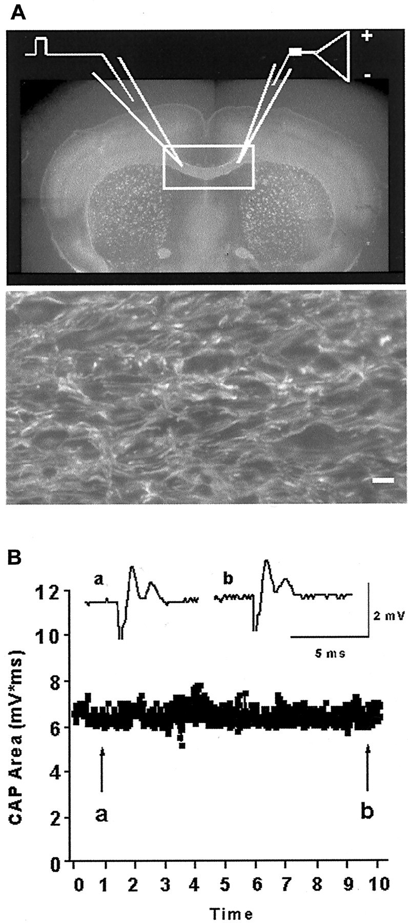
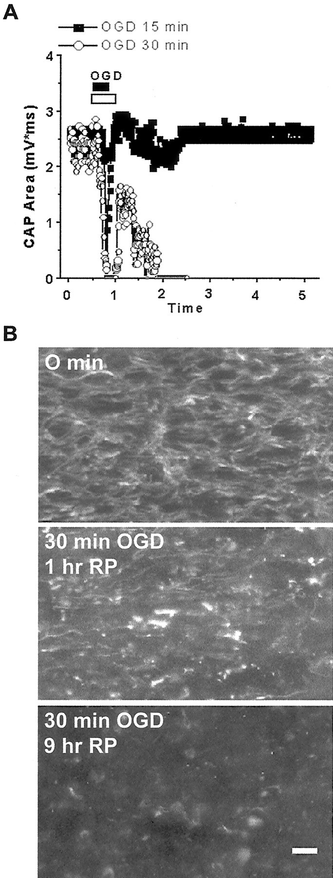
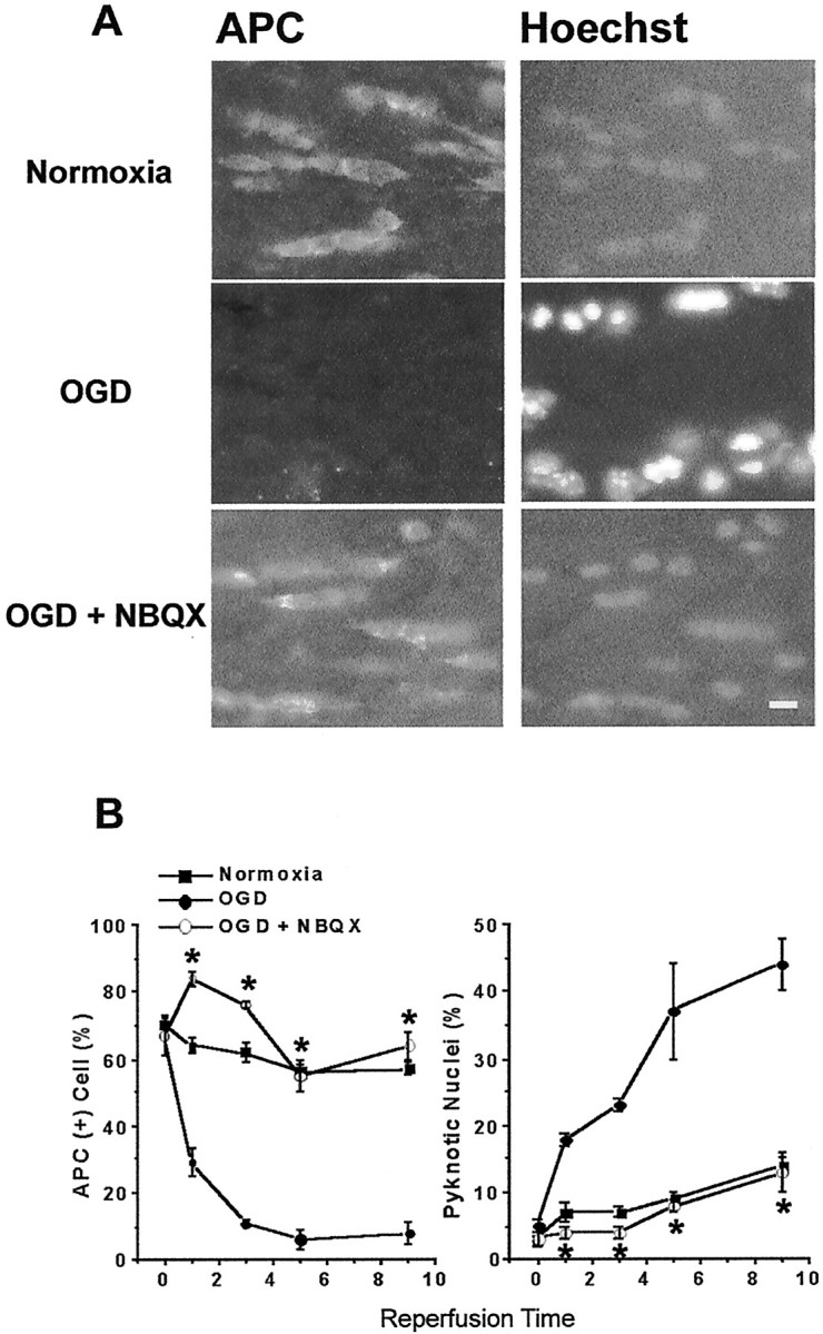
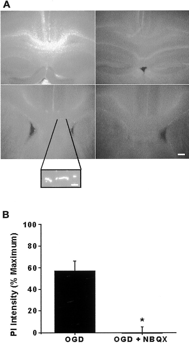
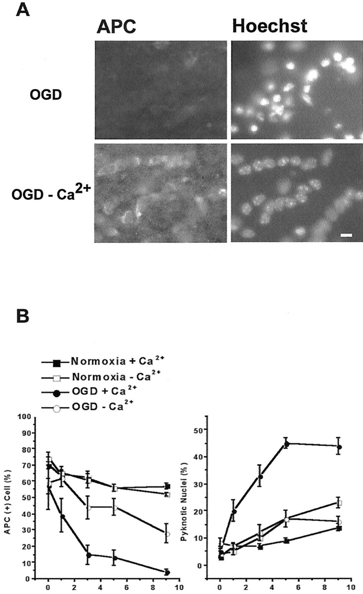
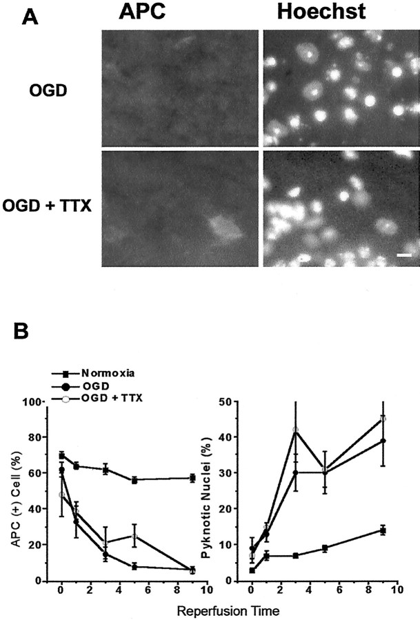
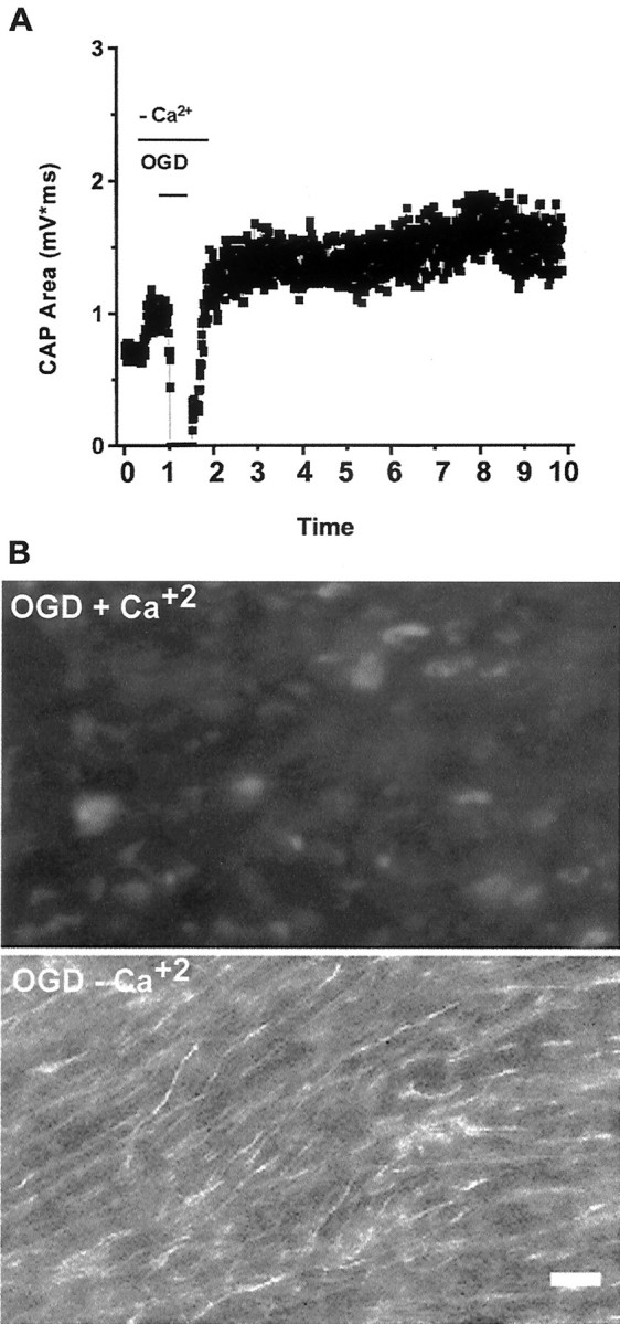
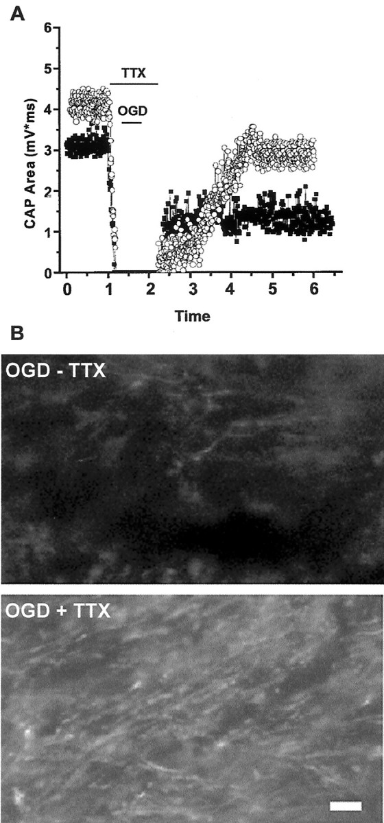
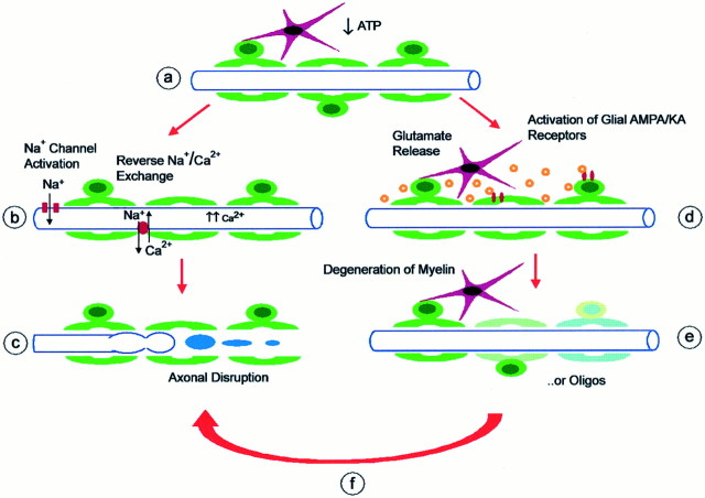
We developed an in situ model to investigate the hypothesis that AMPA/kainate (AMPA/KA) receptor activation contributes to hypoxic-ischemic white matter injury in the adult brain. Acute coronal brain slices, including corpus callosum, were prepared from adult mice. After exposure to transient oxygen and glucose deprivation (OGD), white matter injury was assessed by electrophysiology and immunofluorescence for oligodendrocytes and axonal neurofilaments. White matter cellular components and the stimulus-evoked compound action potential (CAP) remained stable for 12 hr after preparation. OGD for 30 min resulted in an irreversible loss of the CAP as well as structural disruption of axons and subsequent loss of neurofilament immunofluorescence. OGD also caused widespread oligodendrocyte death, demonstrated by the loss of APC labeling and the gain of pyknotic nuclear morphology and propidium iodide labeling. Blockade of AMPA/KA receptors with 30 microm NBQX or the AMPA-selective antagonist 30 microm GYKI 52466 prevented OGD-induced oligodendrocyte death. Oligodendrocytes also were preserved by the removal of Ca(2+), but not by a blockade of voltage-gated Na(+) channels. The protective action of NBQX was still present in isolated corpus callosum slices. CAP areas and axonal structure were preserved by Ca(2+) removal and partially protected by a blockade of voltage-gated Na(+) channels. NBQX prevented OGD-induced CAP loss and preserved axonal structure. These observations highlight convergent pathways leading to hypoxic-ischemic damage of cerebral white matter. In accordance with previous suggestions, the activation of voltage-gated Na(+) channels contributes to axonal damage. Overactivation of glial AMPA/KA receptors leads to oligodendrocyte death and also plays an important role in structural and functional disruption of axons.


Figures

Fig. 1.
Fig. 1.
Immunohistochemical localization of white matter components in brain slices. A, Fluorescence photomicrograph shows oligodendrocytes (APC;green) astrocytes (GFAP;red), and cell nuclei (Hoechst;blue) in the corpus callosum. Intrafascicular oval oligodendrocyte cell bodies and stellate astrocytes are recognized easily and do not overlap. Scale bar, 10 μm. B, Glial cell counts in the corpus callosum are stable over 12 hr in the acute brain slice preparation. Histograms summarize the APC+ (dark gray) and GFAP+ (gray) cell counts in slices from perfusion-fixed, immediately fixed, and normoxic (12 hr) groups. Values represent the mean proportion ± SEM of Hoechst-positive nuclei that express the cell-specific marker. *Significantly different (p < 0.05) from perfusion-fixed conditions. **Significantly different from perfusion-fixed but not immediately fixed conditions.
Fig. 2.
Fig. 2.
Axonal structural and functional assessment in brain slices. A, Placement of recording (right) and stimulation (left) electrodes across the corpus callosum (rectangle) in a perfusion-fixed slice labeled with SMI-31 for neurofilaments.Bottom panel shows higher magnification (60×) of SMI-31-labeled axons from same region. Note parallel linear structure of axons. Scale bar: Top, 125 μm;bottom, 10 μm. B, Compound action potential (CAP) area measurements remain stable over time. Insets are sample traces chosen at the beginning (a) and at the end (b) of a 10 hr recording period.
Fig. 3.
Fig. 3.
The 30 min OGD causes loss of axonal function and structural injury. A, CAP recovers completely after 15 min OGD (filledsquares), but not after 30 min OGD (open circles). B, Immunofluorescence micrograph shows SMI-31 labeling of axons in control (top), 1 hr after 30 min OGD (1 hr RP;middle), and 9 hr after OGD (9 hr RP;bottom). Note linear fibers in control, formation of axonal heads and retraction bulbs 1 hr after OGD, and almost complete loss of SMI-31 labeling 9 hr after OGD. Scale bar, 10 μm.
Fig. 4.
Fig. 4.
AMPA/KA receptor blockade prevents OGD-induced oligodendrocyte death. A, Numerous APC+ oligodendrocytes (left) and their nuclei stained with Hoechst (right) are seen in slices kept normoxic for 12 hr (corresponding to 9 hr of reperfusion time). OGD 30 min (in the presence of MK-801) results in the loss of APC immunoreactivity (middle left panel). Pyknotic nuclei are detected with Hoechst staining by increased fluorescence intensity and smaller nuclei (middle right panel). When AMPA/KA receptors are blocked with 30 μm NBQX, many APC+ cells are observed (bottom left panel), and nuclei are not pyknotic. Scale bar, 10 μm. B, Quantitative effects of NBQX. Plot on the left summarizes APC+oligodendrocyte counts over time in normoxia (filled squares), 30 min OGD (filled circles), and 30 min OGD with NBQX (open circles). On theright, pyknotic cell counts in the same conditions are shown. Note the increase in pyknotic nuclei after OGD; *p < 0.05 compared with OGD at matched time points (two-way ANOVA and Dunnett's post test).
Fig. 5.
Fig. 5.
Propidium iodide (PI) detection of OGD-induced cell death. A, Low-magnification (4× objective) images show PI labeling in the corpus callosum of slices exposed to 360 min OGD (maximal injury) or to wash control for 360 min, 30 min OGD followed by 6 hr normoxia, or 30 min OGD with 30 μm NBQX followed by 6 hr normoxia. PI (0.25 μg/ml) was added to the bath 1 hr before imaging. PI intensity was highest in slices from 360 min OGD and lowest in control and in 30 min OGD with NBQX. Micrographs also include neighboring cortex, hippocampus, and ventricles. Scale bar, 50 μm.Inset, High magnification showing PI-positive oligodendrocyte nuclei. Scale bar, 10 μm. B, Quantification of PI intensity. The 30 min OGD results in 56% increase in PI intensity, which is fully preventable by 30 μmNBQX. Values represent mean fluorescence intensity ± SEM of the white matter region of interest, normalized by subtracting wash control and dividing by 360 min OGD values (=100%). *p < 0.05 (Student's t test).
Fig. 6.
Fig. 6.
OGD-induced oligodendrocyte death depends on extracellular Ca2+. Slices are exposed to 30 min OGD in normal aCSF (containing 2.0 mm Ca2+) or aCSF with no Ca2+ including 200 μmEGTA. Perfusion conditions are maintained for 30 min before and during and 30 min after OGD. A, APC immunofluorescence of slices fixed 9 hr after OGD. Exposing slices to OGD in Ca2+-free media reduces oligodendrocyte death (left) and pyknotic nuclei formation (right) compared with OGD with normal Ca2+. Scale bar, 10 μm. B, Oligodendrocyte cell counts in slices from normoxia (filled squares), Ca2+-free normoxia (open squares), OGD (filledcircles), and Ca2+-free OGD (open circles) are summarized in the plot on the left. Ca2+-free aCSF in normoxic conditions does not alter oligodendrocyte numbers. OGD in Ca2+-free aCSF remarkably reduces OGD-induced oligodendrocyte death. B,Right, Pyknotic nuclei number in slices exposed to Ca2+-free OGD is significantly less than in slices exposed to OGD. Values represent mean ± SEM.
Fig. 7.
Fig. 7.
Blockade of voltage-gated sodium channels does not prevent OGD-induced oligodendrocyte death. Slices are exposed to 10 μm TTX or control aCSF for 30 min before and during and 30 min after OGD. A, APC immunofluorescence of slices fixed 9 hr after OGD. TTX does not reduce oligodendrocyte loss or formation of pyknotic nuclei. Scale bar, 10 μm. B, Plots summarize the number of APC+ cells (left) and pyknotic nuclei (right). TTX does not change counts significantly at any time point, as in Figure6.
Fig. 8.
Fig. 8.
Ca2+ dependence of OGD-induced axonal injury. Slices are exposed to Ca2+-free or control aCSF as described in Figure 6. A, Pretreatment of slices with Ca2+-free aCSF preserves stimulus-evoked CAPs. B, SMI-31 immunofluorescence of slices 9 hr after 30 min OGD with (top) or without (bottom) Ca2+. Removal of Ca2+ results in preservation of axonal function (compare with Fig. 3) and neurofilament labeling. Scale bar, 10 μm.
Fig. 9.
Fig. 9.
Blockade of voltage-gated sodium channels offers partial protection of axonal function and structure. A, CAP recovery in slices exposed to TTX for 90 min, with normoxic perfusion (open circles) or 30 min OGD (filled squares). TTX exposure alone results in complete loss of CAP, which recovers to ∼61% of initial values 5 hr after removal. CAP recovery in slices exposed to OGD + TTX is better than OGD with no drug (see Fig. 3) but does not reach levels of TTX in normoxia. B, Compared with OGD without TTX (top), SMI-31 labeling is preserved partially in slices exposed to OGD + TTX, but some axonal head and retraction bulb formation occurs (bottom).
Fig. 10.
Fig. 10.
AMPA/KA receptor blockade prevents OGD-induced loss of axonal function. Slices are exposed to 30 min OGD in control aCSF (containing 10 μm MK-801) or aCSF with the further addition of 30 μm NBQX. Images show SMI-31 labeling 9 hr after OGD. Top, Blockade of NMDA receptors with MK-801 delays the immediate OGD-induced reduction of CAPs but has no effect on CAP recovery beyond 3 hr (left) and does not preserve SMI-31 labeling (right). Bottom, NBQX prevents acute CAP loss and preserves CAP for 6 hr after OGD. Axonal structure is protected substantially. Scale bar, 10 μm.
Fig. 11.
Fig. 11.
Proposed model for hypoxic–ischemic injury pathways in cerebral white matter. Schematic shows myelinated axon (blue), oligodendrocytes (green), and astrocytes (red).A, Hypoxia, ischemia, or glucose deprivation results in energy depletion and loss of ATP. B, Failure of Na+/K+-ATPase and depolarization leads to opening of noninactivating axonal voltage-gated Na+ channels. Ca2+ enters axons by reversal of Na+/Ca2+ exchange and activation of voltage-gated Ca2+ channels. Action potentials are halted reversibly by loss of ionic gradients.C, Excessive axoplasmic Ca2+ levels trigger destructive pathways, leading to degradation of axonal cytoskeleton and organelles, focal axonal swelling, and eventual interruption of axonal integrity. D, Another effect of energy deprivation is release of glutamate into extracellular space (likely by reversal of Na+-dependent glutamate transport from axons and possibly astrocytes, oligodendrocytes). Glutamate activates ionotropic AMPA/KA receptors on oligodendrocytes (and possibly astrocytes). E, Sustained glutamate receptor activation triggers excitotoxic damage of oligodendrocyte processes (myelin) and subsequent death of oligodendrocytes. Myelin damage might result in conduction delay or block.F, Activation of glial AMPA/KA receptors or oligodendrocyte death triggers further damage to axons under energy-depleted conditions. Mechanisms linking glial glutamate receptors to axonal damage might include release of toxic substances from injured cells, increase in tissue energy use, loss of substrate or trophic support, exposure of protected membrane or ion channels, or failure of glial homeostatic functions. Blockade of stepD by NBQX protects oligodendrocytes and axons, whereas blockade of step B by TTX provides partial protection for axons, but not for oligodendrocytes. Other interactions are not shown. Ion homeostatic failure in axons may be responsible for glutamate release; Na+ removal and a blockade of Na+/Ca2+ exchange likely act on both axons and oligodendrocytes.

References

    1. Adams JH, Graham DI, Gennarelli TA. Head injury in man and experimental animals: neuropathology. Acta Neurochir Suppl (Wien) 1983;32:15–30. - PubMed
    1. Agrawal SK, Fehlings MG. Mechanisms of secondary injury to spinal cord axons in vitro: role of Na+, Na+-K+-ATPase, the Na+–H+ exchanger, and the Na+–Ca2+ exchanger. J Neurosci. 1996;16:545–552. - PMC - PubMed
    1. Agrawal SK, Fehlings MG. Role of NMDA and non-NMDA ionotropic glutamate receptors in traumatic spinal cord axonal injury. J Neurosci. 1997;17:1055–1063. - PMC - PubMed
    1. Anderson CM, Swanson RA. Astrocyte glutamate transport: review of properties, regulation, and physiological functions. Glia. 2000;32:1–14. - PubMed
    1. Baltan Tekkök S, Hyrc KL, Underhill SM, Goldberg MP. Na+/Ca2+ exchange blocker KB-R7943 protects axons and oligodendrocytes during oxygen glucose deprivation. Soc Neurosci Abstr. 2000;26:2065.

Publication types

MeSH terms

LinkOut - more resources