. 2001 Jun 15;21(12):4290-8.
doi: 10.1523/JNEUROSCI.21-12-04290.2001.

Repellent signaling by Slit requires the leucine-rich repeats

Affiliations

Repellent signaling by Slit requires the leucine-rich repeats

R Battye et al. J Neurosci. .

Abstract

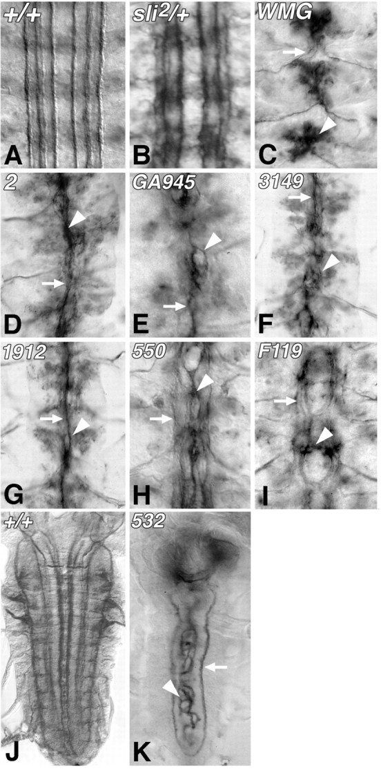
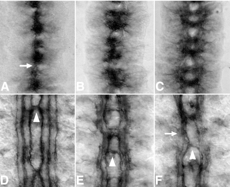
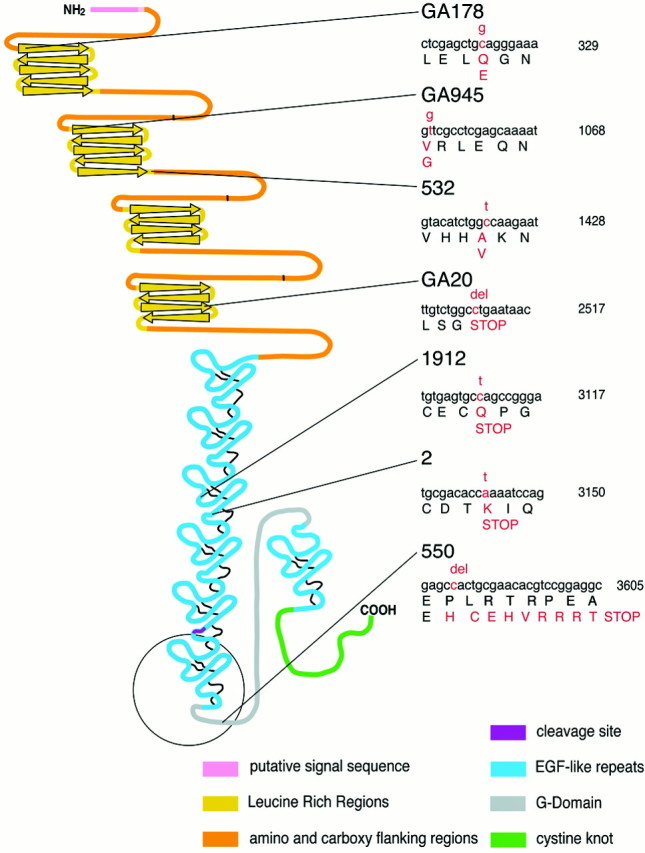
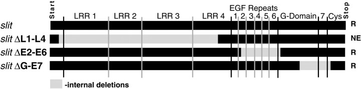
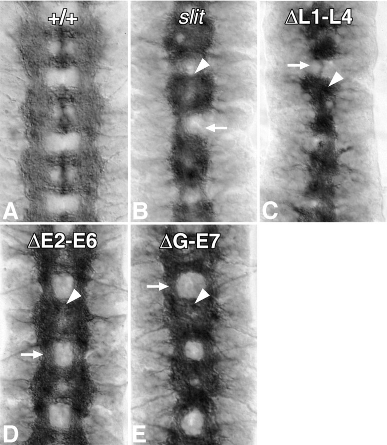
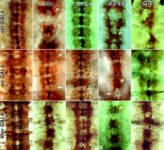
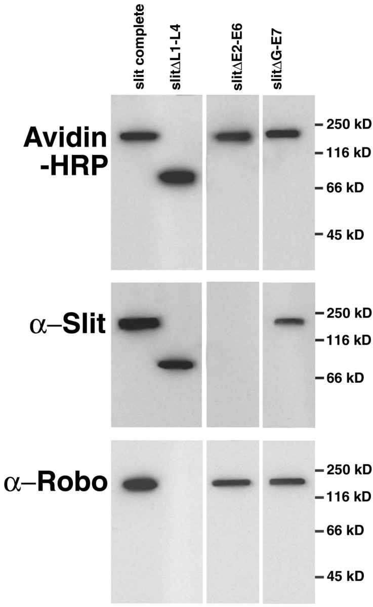
Slit is a repellent axon guidance cue produced by the midline glia in Drosophila that is required to regulate the formation of contralateral projections and the lateral position of longitudinal tracts. Four sequence motifs comprise the structure of Slit: a leucine-rich repeat (LRR), epidermal growth factor-like (EGF) repeats, a laminin-like globular (G)-domain, and a cysteine domain. Here we demonstrate that the LRR is required for repellent signaling and in vitro binding to Robo. Repellent signaling by slit is reduced by point mutations that encode single amino acid changes in the LRR domain. By contrast to the EGF or G-domains, the LRR domain is required in transgenes to affect axon guidance. Finally, we show that the midline repellent receptor, Robo, binds Slit proteins with internal deletions that also retain repellent activity. However, Robo does not bind Slit protein missing the LRR. Taken together, our data demonstrate that Robo binding and repellent signaling by Slit require the LRR region.


Figures

Fig. 1.
Fig. 1.
Midline guidance phenotypes of slitnulls and hypomorphs. In wild-type embryos (A), Fasciclin II labels a bilateral set of three fascicles that run symmetrically along the length of the nerve cord but do not cross the midline or each other. A nerve cord heterozygous forslit2 (B) has slight deviations in each longitudinal fascicle, but individual fascicles remain separated. Fasciclin II labeling axons fuse at the midline in embryos deficient for slit(C) [Df(2R)WMG]. Slit alleles with severe (D,E), moderate (F, G), and mild (H, I) phenotypes are shown. Alleles that do not make immunodetectable Slit,slit2 (D), andslitGA945 (E) cause a complete fusion of longitudinal axons at the midline. Embryos of moderate alleles of slit that generate detectable protein have longitudinal fascicles that crisscross the midline (arrowhead) and have thicker intersegmental connections [slit3149 (F),slit1912 (G)]. Longitudinal axons in the moderate phenotypes still regularly fuse with adjacent fascicles. slit alleles that produced mild phenotypes included slit550(H) andslitF119(I). Mild slit phenotypes have a larger separation between axon fascicles, and contralateral projections across the midline are restricted to the two most medial fascicles. Anterior is at top in these frontal views of stage 16 nerve cords. Numerous Fasciclin II-labeling longitudinal fascicles are found in a wild-type third instar larval nerve cord (J). A second instar slit532 hypomorph of the same age has disorganized medial fascicles that re-cross the midline (K, arrowhead). The most lateral tract is normal (K, arrow). In this and successive Figures, arrows identify intersegmental segments of the longitudinal fascicles, and arrowheadsindicate midline guidance errors.
Fig. 2.
Fig. 2.
Commissure structure and the genetic interaction of slit alleles withrobo1. Embryos homozygous for a severe allele of slit(sli2) lack all commissure structure (A, arrow). For comparison, a wild-type axon tract scaffold labeled with BP102 has a ladder-like morphology (Fig. 5A). Less midline compression of axons and more intersegmental axon projections are seen with moderate [sli1912 (B)] and mild [sli2990(C)] alleles. Separation of commissures (arrow) seen in mild slit mutants is accompanied by greater widening of the ventral nerve cord, resembling arobo-like phenotype. The phenotypic interaction withrobo1 is contrasted with the same alleles. In embryos heterozygous forrobo1 andslit2, deviations are observed in the most medial longitudinal fascicles within a subset of segments (D, arrowhead). The remaining fascicles are typical of embryos heterozygous for eitherslit2 orrobo1. The interactions betweenrobo1 and moderateslit alleles [slit1912(E)] have prominent crossovers (arrowhead). More lateral fascicles are less organized (arrow). A similar pattern is seen in theslit2990/robo1transheterozygotes (F), including the fusion of lateral fascicles (arrow).
Fig. 3.
Fig. 3.
Coding changes of seven slitalleles. Unique changes in the nucleotide and amino acid sequence ofslit alleles (right) are mapped to the structural domain of the Slit protein (left) following the protein motif conventions introduced by Rothberg et al. (1990).
Fig. 4.
Fig. 4.
slit transgenes with internal deletions. The regions of slit deleted in three transgenes are lightly shaded. Letters atright indicate repellent activity. R, Repellent; NE, no effect.
Fig. 5.
Fig. 5.
Restoration of slit function usingslit transgenes with internal deletions. Possible restoration of axon tract architecture was assessed inDrosophila mutant forslit2, also carrying UAS-slit constructs (Fig. 4) expressed underslit1.0-GAL4 regulation. Wild-type expression of Slit in the ventral nerve cord (black) is shown in a BP102-labeled nerve cord (A). The expression of P[UAS-slit complete] was sufficient to partially rescue the slit2 phenotype, demonstrated by greater separation of axons from the midline (B, arrowhead), although intersegmental connections are not restored (B, arrow). A slit transgene lacking the leucine domains ofslit (C) [P(UAS-slitΔL1–L4)] did not rescue theslit2 phenotype. Midline fusion was not suppressed (arrowhead). A slittransgene lacking five EGF repeats, P[UAS-slitΔE2–E6], was able to restore the midline scaffold to near normal conditions, including significant restoration of longitudinal tracts (D). Removal of only the G-domain and final EGF, P[UAS-slitΔG-E7] (E), rescued theslit2 phenotype to a degree similar to slit lacking five EGF repeats (P[UAS-slitΔE2–E6]) (D).
Fig. 6.
Fig. 6.
Ectopic expression of slittransgenes with internal deletions. slit transgenes (Fig. 4) were expressed in three different patterns using specific P[GAL4] drivers in a wild-type background. P[slit1.0-GAL4] (AE) drives expression in the MG, P[eng-GAL4] (FJ) drives ectopic expression at the segmental boundary, and P[elav-GAL4] (KO) directs expression in all neurons. The pattern of ectopic expression is shown in A,F, and K, where each P[GAL4] driver was crossed to P[UAS-tau-LacZ] and embryos were labeled with BP102 (black) and α−β-gal (brown). In the remaining panels, BP102 (brown) and Slit immunolabeling (black) are visualized. Expression of the completeslit cDNA (B, G,L) disrupted both longitudinal (arrows) and commissural tracts. Some axons misproject laterally (B, asterisk). Ectopic expression of P[UAS-slit complete] in all neurons (L) displaced axon tracts toward the midline. Expression of a slit transgene that lacks the leucine domains (C, H,M) did not significantly alter axon tract organization. Expression of a slit transgene lacking EGF repeats 2 through 6 (P[UAS-slitΔE2-E6]) significantly altered axon tract organization (D,I, N). Intersegmental expression of P[UAS-slitΔE2-E6] resulted in a displacement of axons toward the midline (I, arrowhead) and breaks in the longitudinal tracts (arrow). Expression of P[UAS-slitΔE2-E6] in all neurons also eliminates most intersegmental axons (N,arrow). This construct was not detected by Slit antibody, and only native Slit labeling is seen. Expression of aslit transgene lacking the G-domain and the seventh EGF repeat (E, J,O) results in effects similar to expression of the entire construct. Intersegmental expression of P[UAS-slitΔG-E7] resulted in a number of breaks in longitudinal fascicles (J, arrow) and poorly defined anterior and posterior commissures (J,arrowhead). Expression of P[UAS-slitΔG-E7] in all neurons (O) has displaced axons toward the midline (arrowhead).
Fig. 7.
Fig. 7.
In vitro binding of truncated Slit to Robo. Slit transgenes, both complete and containing internal deletions (Fig. 4), were in vitro translated incorporating biotin-labeled lysine and then bound to avidin Agarose columns. In vitro translated Robo incubated with each column, washed, and eluted with n-biotin. Thirty percent of the extraction from each column was immunoblotted as indicated. Avidin-HRP visualized with chemoluminescence shows that each of the Slit constructs bound and was then eluted from the avidin columns. The Slit antibody detects all constructs except those that delete the EGF2–EGF6 region, to which the antibody binds. The immunoblot for Robo indicates that Slit complete, SlitΔE2-E6, and SlitΔG are capable of binding Robo; however, Slit protein with internal deletions of any or all of the LRR fail to demonstrate any detectable roboantigen.

Similar articles

Cited by

References

    1. Battye R, Stevens A, Jacobs JR. Axon repulsion from the midline of the Drosophila CNS requires slit function. Development. 1999;126:2475–2481. - PubMed
    1. Brand AH, Perrimon N. Targeted gene expression as a means of altering cell fates and generating dominant phenotypes. Development. 1993;118:401–415. - PubMed
    1. Brose K, Bland KS, Wang K-H, Arnott D, Henzel W, Goodman CS, Tessier-Lavigne M, Kidd T. Evolutionary conservation of the repulsive guidance function of Slit proteins and their interactions with robo receptors. Cell. 1999;96:795–806. - PubMed
    1. Cornish T, Chi J, Johnson S, Lu Y, Campanelli JT. Globular domains of agrin are functional units that collaborate to induce acetylcholine receptor clustering. J Cell Sci. 1999;112:1213–1223. - PubMed
    1. Gay NJ, Packman LC, Weldon MA, Barna JC. A leucine-rich repeat peptide derived from the Drosophila Toll receptor forms extended filaments with a beta-sheet structure. FEBS Lett. 1991;291:87–91. - PubMed

Publication types

MeSH terms

LinkOut - more resources