The role of nitric oxide in development of topographic precision in the retinotectal projection of chick
- PMID: 11404417
- PMCID: PMC6762761
- DOI: 10.1523/JNEUROSCI.21-12-04318.2001
The role of nitric oxide in development of topographic precision in the retinotectal projection of chick
Abstract
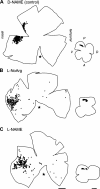
The axonal projection from the retina to the tectum exhibits a precise topographic order in the mature chick such that neighboring ganglion cells send axons to neighboring termination zones in the contralateral tectum. The initial pattern formed during development is much less organized and is refined to the adult pattern during a discrete period of development. Refinement includes elimination of radically aberrant projections, such as those from the temporal side of the retina to posterior regions of the tectum, as well as a more subtle improvement in the topographic precision of the projection. The enzyme that synthesizes nitric oxide is expressed at high levels in the tectum during the developmental period in which the topography improves. Pharmacological blockade of nitric oxide synthesis during this period prevented elimination of topographically inappropriate retinotectal projections in a dose-dependent manner. This effect could not be duplicated by treatment of embryos with a vasoconstrictor, indicating that vascular changes were not a factor. These results show that nitric oxide is involved in refinement of the topography of the retinotectal projection as well as in other aspects of refinement of this projection in developing chick.
Figures


Similar articles
-
NMDA receptor-mediated refinement of a transient retinotectal projection during development requires nitric oxide.J Neurosci. 1999 Jan 1;19(1):229-35. doi: 10.1523/JNEUROSCI.19-01-00229.1999. J Neurosci. 1999. PMID: 9870953 Free PMC article.
-
Involvement of nitric oxide in the elimination of a transient retinotectal projection in development.Science. 1994 Sep 9;265(5178):1593-6. doi: 10.1126/science.7521541. Science. 1994. PMID: 7521541
-
Mechanisms involved in development of retinotectal connections: roles of Eph receptor tyrosine kinases, NMDA receptors and nitric oxide.Prog Brain Res. 1998;118:115-31. doi: 10.1016/s0079-6123(08)63204-5. Prog Brain Res. 1998. PMID: 9932438 Review.
-
Topographic-specific axon branching controlled by ephrin-As is the critical event in retinotectal map development.J Neurosci. 2001 Nov 1;21(21):8548-63. doi: 10.1523/JNEUROSCI.21-21-08548.2001. J Neurosci. 2001. PMID: 11606643 Free PMC article.
-
Development of the visual system of the chick. II. Mechanisms of axonal guidance.Brain Res Brain Res Rev. 2001 Jul;35(3):205-45. doi: 10.1016/s0165-0173(01)00049-2. Brain Res Brain Res Rev. 2001. PMID: 11423155 Review.
Cited by
-
Rac1-mediated endocytosis during ephrin-A2- and semaphorin 3A-induced growth cone collapse.J Neurosci. 2002 Jul 15;22(14):6019-28. doi: 10.1523/JNEUROSCI.22-14-06019.2002. J Neurosci. 2002. PMID: 12122063 Free PMC article.
-
Nitric oxide as a putative retinal axon pathfinding and target recognition cue in Xenopus laevis.Impulse (Columbia). 2011 Jan 1;2010:1-12. Impulse (Columbia). 2011. PMID: 21847432 Free PMC article.
-
Chemical signaling in the developing avian retina: Focus on cyclic AMP and AKT-dependent pathways.Front Cell Dev Biol. 2022 Dec 9;10:1058925. doi: 10.3389/fcell.2022.1058925. eCollection 2022. Front Cell Dev Biol. 2022. PMID: 36568967 Free PMC article. Review.
References
-
- Arancio O, Kiebler M, Lee C, Lev-Ram V, Tsien R, Kandel E, Hawkins R. Nitric oxide acts directly in the presynaptic neuron to produce long-term potentiation in cultured hippocampal neurons. Cell. 1996;87:1025–1035. - PubMed
-
- Boulanger L, Poo MM. Presynaptic depolarization facilitates neurotrophin-induced synaptic potentiation. Nat Neurosci. 1999;2:346–351. - PubMed
-
- Campello-Costa P, Fosse AM, Ribeiro JC, Paes-de-Carvalho R, Serfaty CA. Acute blockade of nitric oxide synthesis induces disorganization and amplifies lesion-induced plasticity in the rat retinotectal projection. J Neurobiol. 2000;44:371–381. - PubMed
-
- Chen Y, Naito J. A quantitative analysis of cells in the ganglion cell layer of the chick retina. Brain Behav Evol. 1999;53:75–86. - PubMed
Publication types
MeSH terms
Substances
Grants and funding
LinkOut - more resources
Full Text Sources
