

doi: 10.1073/pnas.131179698.
Epub 2001 Jun 12.
Crystal structure of an anticoagulant protein in complex with the Gla domain of factor X
Affiliations
- PMID: 11404471
- PMCID: PMC34651
- DOI: 10.1073/pnas.131179698
Item in Clipboard
Crystal structure of an anticoagulant protein in complex with the Gla domain of factor X
Proc Natl Acad Sci U S A.
.
Abstract
The gamma-carboxyglutamic acid (Gla) domain of blood coagulation factors is responsible for Ca2+-dependent phospholipid membrane binding. Factor X-binding protein (X-bp), an anticoagulant protein from snake venom, specifically binds to the Gla domain of factor X. The crystal structure of X-bp in complex with the Gla domain peptide of factor X at 2.3-A resolution showed that the anticoagulation is based on the fact that two patches of the Gla domain essential for membrane binding are buried in the complex formation. The Gla domain thus is expected to be a new target of anticoagulant drugs, and X-bp provides a basis for designing them. This structure also provides a membrane-bound model of factor X.
Figures
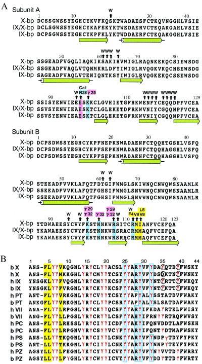
Amino acid sequence alignments. (A) Anticoagulant proteins from snake venoms. Black arrows above the residues of X-bp point to amino acid residues of XGD1-44, Ca-1, or water molecules (W) with which they interact. Positively charged, negatively charged, and hydrophobic residues involved in those interactions are colored in blue, pink, and yellow, respectively. The numbering and secondary structure elements also are shown. (B) The Gla domains of vitamin K-dependent proteins (X, factor X; IX, factor IX; PT, prothrombin; VII, factor VII; PC, protein C; PS, protein S; PZ, protein Z) from human (h) and bovine (b). The numbering at the top refers to the bX sequence. Gla residues are denoted by γ and colored in red. Blue and yellow columns show hydrophilic and hydrophobic patches, respectively. Residues coordinating to the eighth Ca2+ ion are indicated in the circles.
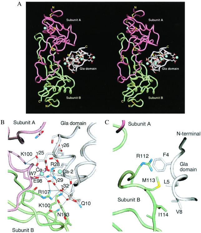
Overall structure of X-bp and XGD1-44 complex. (A) Stereoview of ribbon model viewed perpendicular to the pseudodyad of the molecule, showing the interface between X-bp and XGD1-44. The subunits A and B of X-bp are magenta and green. XGD1-44 is white. The side chain of Gla residues and disulfide bonds are displayed. The bound Ca2+ ions are denoted by blue spheres. (B) Same view as in A, but molecular detail of interaction between the hydrophilic patch of XGD1-44 and positively charged X-bp, and a bridging Ca2+. (C) Same view as in B, but between the N terminus hydrophobic patch of XGD1-44 and the C terminus of subunit B of X-bp.
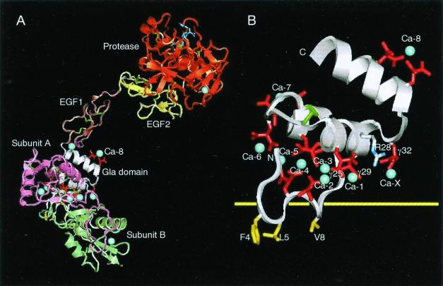
Model of factor Xa bound to X-bp and XGD1-44 bound to membrane. (A) Factor Xa bound to X-bp. The Gla residues are in red, bound Ca2+ ions in blue (only labeled is Ca-8, which is identified in the present study), and disulfide bonds in green. The small molecule (dark blue) bound to the active site of the protease domain is the FX-2212a inhibitor (17). (B) Putative membrane-binding surface of XGD1-44. Same view as in A, but the scale of the figure is magnified for clarity. The hydrophobic patch includes Phe4, Leu5, and Val8, and hydrophilic patch includes Arg28, Gla25, Gla29, Gla32, and Ca-1 as a bridging Ca2+, which are on either side of the yellow horizontal line of the putative membrane surface. Ca-X is a putative Ca2+ ion that is taken in as another bridging Ca2+.
References
-
- Mann K G. Thromb Haemostasis. 1999;82:165–174. - PubMed
-
- Leung D, Abbenante G, Fairlie D P. J Med Chem. 2000;43:305–341. - PubMed
-
- Dennis M S, Elgenbrot C, Skelton N J, Ultsch M H, Santell L, Dwyer M A, O'Connell M P, Lazarus R A. Nature (London) 2000;404:465–470. - PubMed
-
- Atoda H, Hyuga M, Morita T. J Biol Chem. 1991;266:14903–14911. - PubMed
-
- Atoda H, Ishikawa M, Yoshihara E, Sekiya F, Morita T. J Biochem. 1995;118:965–973. - PubMed
Publication types
MeSH terms
Substances
Associated data
- Actions
LinkOut - more resources
Full Text Sources
Other Literature Sources
Miscellaneous
