. 2001 Jun 15;20(12):3046-55.
doi: 10.1093/emboj/20.12.3046.

EndoS, a novel secreted protein from Streptococcus pyogenes with endoglycosidase activity on human IgG

Affiliations

EndoS, a novel secreted protein from Streptococcus pyogenes with endoglycosidase activity on human IgG

M Collin et al. EMBO J. .

Abstract

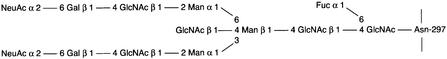
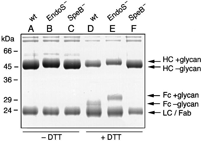
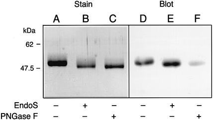
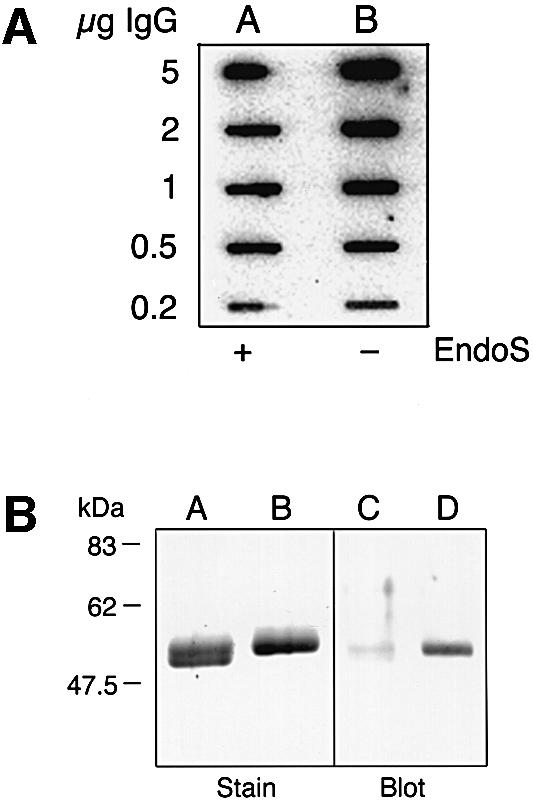
Streptococcus pyogenes is an important human pathogen that selectively interacts with proteins involved in the humoral defense system, such as immunoglobulins and complement factors. In this report we show that S.pyogenes has the ability to hydrolyze the chitobiose core of the asparagine-linked glycan on immuno globulin G (IgG) when bacteria are grown in the presence of human plasma. This activity is associated with the secretion of a novel 108 kDa protein denoted EndoS. EndoS has endoglycosidase activity on purified soluble IgG as well as IgG bound to the bacterial surface. EndoS is required for the activity on IgG, as an isogenic EndoS mutant could not hydrolyze the glycan on IgG. In addition, we show that the secreted streptococcal cysteine proteinase SpeB cleaves IgG in the hinge region in a papain-like manner. This is the first example of an endoglycosidase produced by a bacterial pathogen that selectively hydrolyzes human IgG, and reveals a novel mechanism which may contribute to S.pyogenes pathogenesis.


Figures

None
Fig. 1. Structure of the fully substituted complex biantennary type oligosaccharide attached to Asn297 of the IgG γ-chain. GlcNAc, N-acetylglucosamine; Fuc, fucose; Man, mannose; Gal, galactose; NeuAc, sialic acid.
None
Fig. 2. Identification of a prominent, high molecular weight, secreted protein from S.pyogenes (indicated by an arrow). Proteins present in bacterial culture supernatants were separated by two identical 10% SDS–PAGE, one stained with Coomassie Blue (Stain), the other blotted to a membrane and detected with polyclonal rabbit EndoS antiserum (Blot). Lanes A and C, AP1 (wild type); lanes B and D, MC14 (EndoS).
None
Fig. 3. EndoS is similar to EndoF2. Sequence alignment of EndoS from S.pyogenes strain AP1 and EndoF2 from F.meningosepticum. The N-terminal sequence of the protein band isolated from AP1 culture supernatant is underlined (EEKTVQVQ). The chitinase motif is indicated in bold (LDGLDVDVE) and the essential glutamic acid for activity is marked with an asterisk. The consensus sequence is shown under the alignment and similarities are indicated with dots.
None
Fig. 4. (A) A schematic drawing of the strategy for mutagenesis of the ndoS gene in AP1 bacteria. An internal 534 bp fragment from the ndoS gene was cloned in the suicide plasmid pFW13, generating pMC11. This plasmid was electroporated into AP1 for homologous recombination. (B) PCR analysis of purified chromosomal DNA from AP1 and MC14. The vector- and ndoS-derived primers used are indicated with roman numerals I–IV in (A) and (B).
None
Fig. 5. EndoS has glycosidase activity on IgG in human plasma. Streptococcus pyogenes AP1 and MC14 were grown in diluted CM (12.5%) supplemented with 1% human plasma. After overnight growth, culture supernatants were analyzed by 10% SDS–PAGE. Lane A, proteins from growth with AP1 (wild type); lane B, proteins from growth with MC14 (EndoS); lane C, medium alone. The 27 and the 31 kDa fragments of the γ-chains are indicated with arrows on the right.
None
Fig. 6. Removal of Fc glycans by EndoS and cleavage of the hinge region by SpeB. SDS–PAGE analysis of purified human IgG incubated with supernatants from AP1 (wt), MC14 (EndoS) and AL1 (SpeB). Samples were incubated under non-reducing (–DTT) or reducing (+DTT) conditions, as indicated. Lanes A and D, wild type (wt); lanes B and E, EndoS; lanes C and F, SpeB. Arrows on the right indicate the position of heavy chains with glycan (HC +glycan) or without glycan (HC –glycan), Fc fragment with glycan (Fc +glycan) or without glycan (Fc –glycan), light chains (LC) and Fab fragment of heavy chains (Fab).
None
Fig. 7. SDS–PAGE and lectin blot analysis of IgG incubated with and without streptococcal or recombinant EndoS and separated by 10% SDS–PAGE. Gels were analyzed by Coomassie Blue staining (Stain) or by blotting to a membrane that was probed with GNL lectin (Blot). (A) Lanes A and C, IgG incubated with streptococcal EndoS; lanes B and D, IgG incubated without EndoS. (B) IgG incubated with extracts from E.coli expressing EndoS. Lanes A and C, induced cells (+IPTG); lanes B and D, non-induced cells (–IPTG). The arrow on the left indicates the position of recombinant EndoS.
None
Fig. 8. EndoS is an endoglycosidase. Purified human IgG was incubated with either EndoS or PNGase F (PNGase F), separated by 10% SDS–PAGE. Gels were analyzed by Coomassie Blue staining (Stain) or by blotting to a membrane that was probed with UEA-I lectin (Blot). Lanes A and D, untreated IgG; lanes B and E, EndoS-treated IgG; lanes C and F, PNGase F-treated IgG.
None
Fig. 9. (A) Ligand blot analysis of protein H binding to glycosylated or EndoS-treated IgG. Purified IgG was incubated with culture supernatant with or without EndoS, applied to a membrane in serial dilutions, and probed with125I-labeled protein H. Lane A, IgG incubated with EndoS; lane B, IgG incubated without EndoS. (B) EndoS has endoglycosidase activity on IgG when bound to the bacterial surface. SDS–PAGE and GNL lectin blot analysis of IgG, bound to the bacterial surface via protein H, treated with or without EndoS prior to elution at low pH. Lanes A and C, surface-bound IgG incubated in the presence of EndoS; lanes B and D, surface-bound IgG incubated without EndoS.

References

    1. Åkesson P., Cooney,J., Kishimoto,F. and Björck,L. (1990) Protein H—a novel IgG binding bacterial protein. Mol. Immunol., 27, 523–531. - PubMed
    1. Alexander S. and Elder,J.H. (1989) Endoglycosidases from Flavo bacterium meningosepticum: application to biological problems. Methods Enzymol., 179, 505–518. - PubMed
    1. Altschul S.F., Gish,W., Miller,W., Myers,E.W. and Lipman,D.J. (1990) Basic local alignment search tool. J. Mol. Biol., 215, 403–410. - PubMed
    1. Axford J.S., Mackenzie,L., Lydyard,P.M., Hay,F.C., Isenberg,D.A. and Roitt,I.M. (1987) Reduced B-cell galactosyltransferase activity in rheumatoid arthritis. Lancet, 2, 1486–1488. - PubMed
    1. Baenziger J. and Kornfeld,S. (1974) Structure of the carbohydrate units of IgA1 immunoglobulin. I. Composition, glycopeptide isolation and structure of the asparagine-linked oligosaccharide units. J. Biol. Chem., 249, 7260–7269. - PubMed

Publication types