. 2001 Jun 15;20(12):3101-13.
doi: 10.1093/emboj/20.12.3101.

Direct cleavage of the human DNA fragmentation factor-45 by granzyme B induces caspase-activated DNase release and DNA fragmentation

Affiliations

Direct cleavage of the human DNA fragmentation factor-45 by granzyme B induces caspase-activated DNase release and DNA fragmentation

E Sharif-Askari et al. EMBO J. .

Abstract

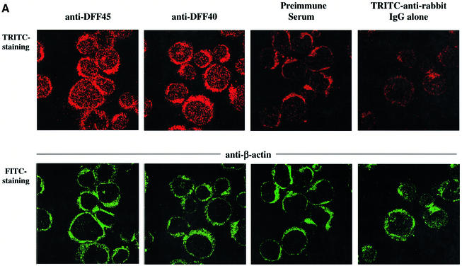
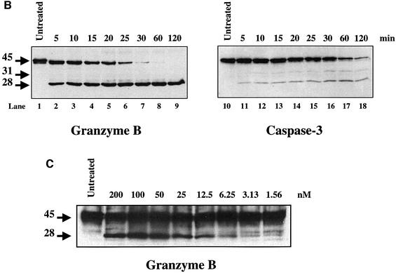
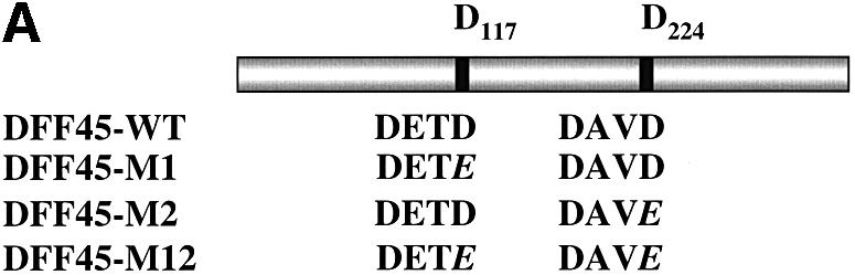
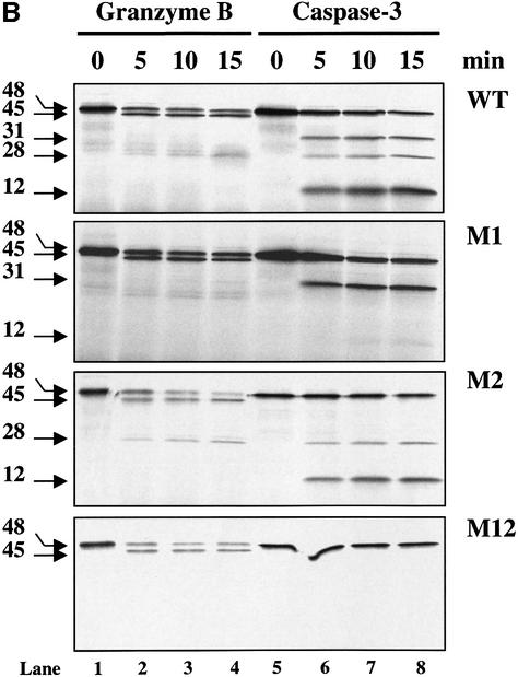
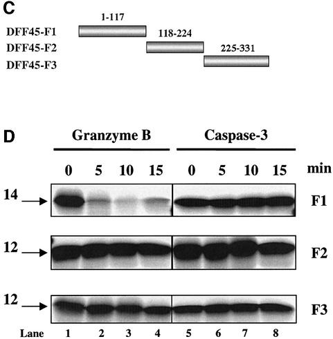
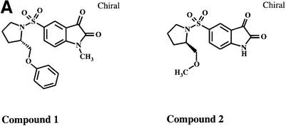
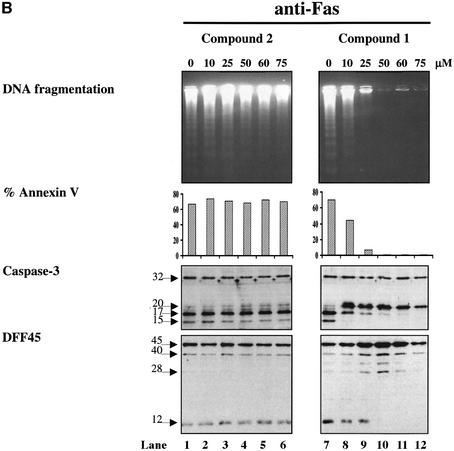
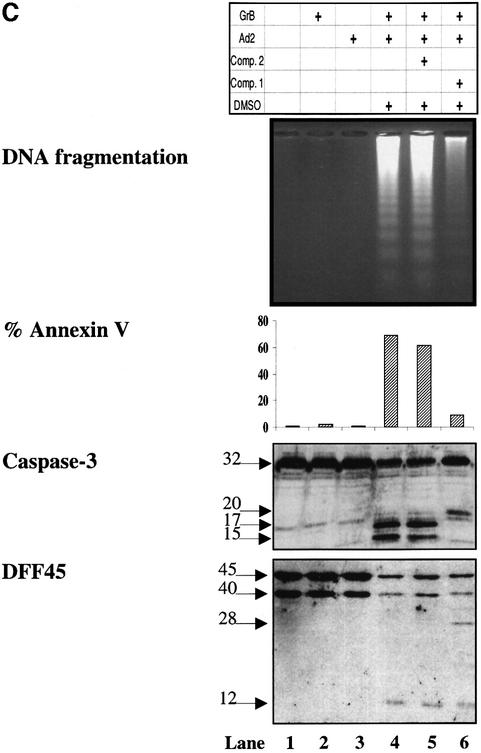
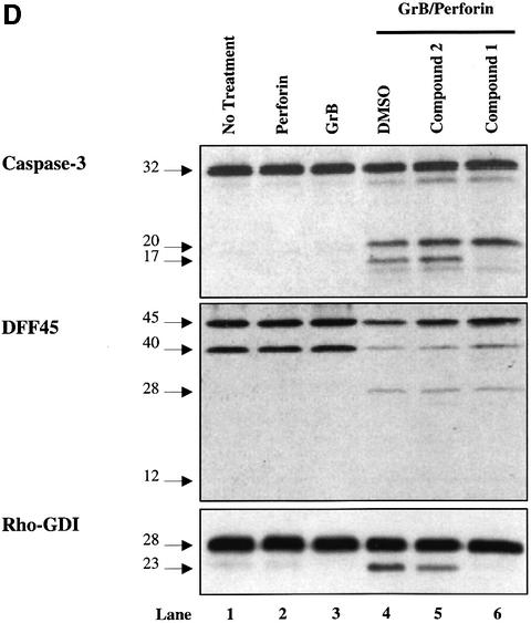
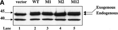
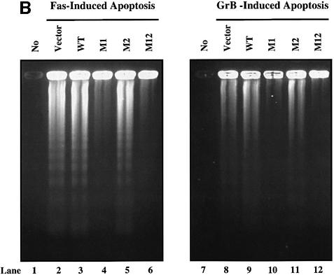
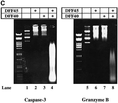
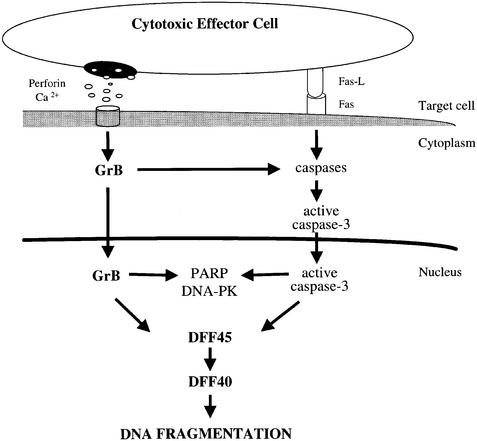
The protease granzyme B (GrB) plays a key role in the cytocidal activity during cytotoxic T lymphocyte (CTL)-mediated programmed cell death. Multiple caspases have been identified as direct substrates for GrB, suggesting that the activation of caspases constitutes an important event during CTL-induced cell death. However, recent studies have provided evidence for caspase-independent pathway(s) during CTL-mediated apoptosis. In this study, we demonstrate caspase-independent and direct cleavage of the 45 kDa unit of DNA fragmentation factor (DFF45) by GrB both in vitro and in vivo. Using a novel and selective caspase-3 inhibitor, we show the ability of GrB to process DFF45 directly and mediate DNA fragmentation in the absence of caspase-3 activity. Furthermore, studies with DFF45 mutants reveal that both caspase-3 and GrB share a common cleavage site, which is necessary and sufficient to induce DNA fragmentation in target cells during apoptosis. Together, our data suggest that CTLs possess alternative mechanism(s) for inducing DNA fragmentation without the requirement for caspases.


Figures

None
Fig. 1. Nuclear compartmentalization and efficient in vitro processing of DFF45 protein by GrB. (A) Double labeling confocal immunofluorescence microscopy of Jurkat cells. Jurkat cells were stained by anti-β-actin monoclonal antibodies and FITC-coupled anti-mouse IgG. DFF45 and DFF40 were detected by using either anti-DFF45 or anti-DFF40 rabbit polyclonal antibodies, respectively, and TRITC-coupled anti-rabbit IgG as described in Materials and methods. Controls were performed by labeling cells with either the secondary TRITC-coupled anti-rabbit IgG alone, or with pre-immune rabbit serum. Each picture is representative of at least 10 fields of two independent experiments at a magnification of 1000×. (B) Time-dependent cleavage of DFF45 by GrB. rDFF45 (12 ng) was incubated for the times indicated with 1 U of GrB (lanes 1–9) or with 10 U of caspase-3 (lanes 10–18) at 37°C. Reactions were stopped by adding Laemmli sample buffer and analyzed by western blotting with anti-DFF45 Ab. Results are representative of three separate experiments. (C) Dose-dependent cleavage of DFF45 by GrB. Purified rDFF45 (12 ng) was incubated with different concentrations of GrB at 37°C for 20 min and subjected to western blot analysis using anti-DFF45 antiserum.
None
Fig. 2. GrB shares the DETD117↓ cleavage site with caspase-3 and exhibits an additional specificity at VTGD6↓ on DFF45. (A) Schematic diagram of wild-type DFF45 (DFF45-WT) and caspase-3 cleavage site mutants (DFF45-M1, -M2, -M12). Overlap PCR was used to mutate aspartic acid (D) residues of the first (D117), second (D224) or both caspase-3 cleavage sites to glutamic acid (E). Caspase-3 cleavage sites are indicated. (B) Radiolabeled His-tagged DFF45 proteins were incubated in the presence of 1 U of GrB (lanes 1–4) or 10 U of caspase-3 (lanes 5–8) for the indicated time intervals, and the reactions were analyzed by autoradiography. Incubation of DFF45-WT or its mutants with GrB or caspase-3 showed that GrB shares the D117 cleavage site with caspase-3 and exhibits additional proteolytic activity. These results are representative of three separate experiments. (C) Identification of the additional GrB cleavage site on DFF45. cDNAs representing DFF45 fragments (DFF45-F1, -F2 and -F3 corresponding to amino acids 1–117, 118–224 and 225–334, respectively) were cloned, and radiolabeled proteins were generated as described in Materials and methods. (D) Radiolabeled proteins were treated as in (B). Only DFF45-F1, corresponding to the N-terminus (1–117), was cleaved by GrB. These results are representative of two separate experiments.
None
Fig. 3. GrB induces DNA fragmentation in the absence of caspase-3 activation. (A) Structures of the caspase-3/-7-specific inhibitor (compound 1) and a 600-fold less active analog (compound 2). (B) Compound 1 blocks Fas-mediated apoptosis in a dose-dependent fashion. Jurkat cells were pre-treated with different concentrations of compound 1 (lanes 7–12) or compound 2 (lanes 1–6). Apoptosis was induced by cross-linking cell surface Fas receptors using mAb to Fas (M3, 20 µg/ml). Abrogation of Fas-induced apoptosis by compound 1 was monitored by DNA fragmentation, annexin V externalization, caspase-3 cleavage and DFF processing. No effect was noticed for compound 2 even at the highest concentration used (lanes 1–6). (C) DFF cleavage and DNA fragmentation occur in the absence of caspase-3 activation. Jurkat cells (1 × 10⁶ cells per well) were pre-treated with 75 µM compound 1 or compound 2. Apoptosis by GrB/Ad2 was induced as described in Materials and methods. Cell death was assayed as in (B). Pre-treatment of the cells with the caspase-3-specific inhibitor compound 1 did not prevent DFF processing and DNA fragmentation induced by GrB (lane 6). Pre-treatment of cells with the caspase-3-specific inhibitor compound 1 (lane 6), but not its analog compound 2 (lane 5), or DMSO alone (lane 4) resulted in a significant decrease in annexin V externalization. Untreated Jurkat cells (lane 1) and Jurkat cells treated with GrB or Ad2 alone (lanes 2 and 3, respectively) showed no significant sign of apoptosis. These results are representative of two separate experiments. (D) Jurkat cells (1 × 10⁵ cells per well) were pre-treated with 75 µM compound 1 or compound 2, and apoptosis was induced by recombinant GrB/perforin as described in Materials and methods. Cell death was assayed by 51Cr release. Pre-treatment with compound 1, but not compound 2, or DMSO blocked further processing of caspase-3 to the p17 active form (lanes 6, 4 and 5, respectively, upper panel) and processing of caspase-mediated cleavage of Rho-GDI substrate (lanes 6, 4 and 5, respectively, lower panel). Blockage of caspase-3 activity had no effect on DFF processing (lane 6, middle panel). Untreated Jurkat cells (lane 1) and those treated with perforin or GrB alone (lanes 2 and 3, respectively) showed no significant sign of apoptosis. These results are representative of three separate experiments.
None
Fig. 4. Cleavage of DFF45 at residue D117 is necessary and sufficient to induce DNA fragmentation. (A) Jurkat cells were stably transfected with SRα Neo/DFF45-WT, -M1, -M2 or -M12 (lanes 2–5, respectively). The empty SRα Neo vector was transfected in Jurkat cells and used as a control (lane 1). Expression of the transfected (exogenous) and native (endogenous) DFF45 is indicated. (B) Stably transfected Jurkat cells were triggered to undergo apoptosis by either Fas (lanes 2–6) or GrB/Ad2 (lanes 8–12) for 5 h as described in Materials and methods. Following the reaction, fragmented DNA was extracted and analyzed on a 2% agarose gel. Mutation of residue D117 completely abolished DNA fragmentation following GrB- (lane 10) or Fas (lane 4)-induced apoptosis. The results are representative of three separate experiments. (C) Direct cleavage of DFF45 by GrB triggers CAD nuclease activity in a cell-free system. The human CAD was expressed in the in vitro transcription and translation system in the absence (lanes 3 and 7) or presence (lanes 4 and 8) of 160 ng of rDFF45 as described in Materials and methods. CAD nuclease activity using plasmid DNA as a substrate was determined in the presence of 0.12 µM GrB (lanes 5–8) or 0.2 µM caspase-3 (lanes 1–4). Samples containing plasmid alone (lanes 1 and 5) or rDFF45 alone (lanes 2 and 6) were subjected to a similar procedure in the presence of GrB (lanes 5 and 6) or caspase-3 (lanes 1 and 2). The results are representative of two separate experiments.
None
Fig. 5. Mechanism of GrB-induced DNA fragmentation in the absence of caspase-3 activity. Following the induction of cellular cytotoxicity, GrB enters target cells and becomes activated in the presence of perforin. Rapid translocation of GrB from the cytoplasm to the nucleus leads to nuclear substrate cleavage. Direct cleavage of DFF45 by GrB induces DNA fragmentation via bypassing of caspase activation. GrB-induced caspase activity will serve to augment the apoptotic process.

References

    1. Alam A., Braun,M.Y., Hartgers,F., Lesage,S., Cohen,L., Hugo,P., Denis,F. and Sekaly,R.P. (1997) Specific activation of the cysteine protease CPP32 during the negative selection of T cells in the thymus. J. Exp. Med., 186, 1503–1512. - PMC - PubMed
    1. Alam A., Cohen,L.Y., Aouad,S. and Sekaly,R.P. (1999) Early activation of caspases during T lymphocyte stimulation results in selective substrate cleavage in nonapoptotic cells. J. Exp. Med., 190, 1879–1890. - PMC - PubMed
    1. Andrade F., Roy,S., Nicholson,D., Thornberry,N., Rosen,A. and Casciola-Rosen,L. (1998) Granzyme B directly and efficiently cleaves several downstream caspase substrates: implications for CTL-induced apoptosis. Immunity, 8, 451–460. - PubMed
    1. Atkinson E.A., Barry,M., Darmon,A.J., Shostak,I., Turner,P.C., Moyer,R.W. and Bleackley,R.C. (1998) Cytotoxic T lymphocyte-assisted suicide. Caspase 3 activation is primarily the result of the direct action of granzyme B. J. Biol. Chem., 273, 21261–21266. - PubMed
    1. Beidler D.R., Tewari,M., Friesen,P.D., Poirier,G. and Dixit,V.M. (1995) The baculovirus p35 protein inhibits Fas- and tumor necrosis factor-induced apoptosis. J. Biol. Chem., 270, 16526–16528. - PubMed

Publication types