. 2001 Jun 15;20(12):3272-81.
doi: 10.1093/emboj/20.12.3272.

Role of the non-homologous DNA end joining pathway in the early steps of retroviral infection

Affiliations

Role of the non-homologous DNA end joining pathway in the early steps of retroviral infection

L Li et al. EMBO J. .

Abstract

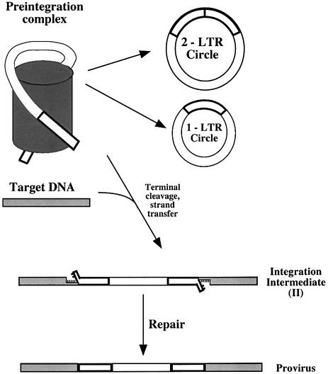
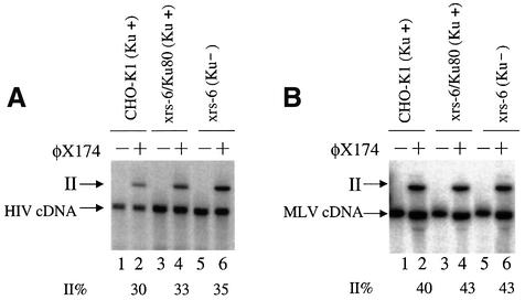
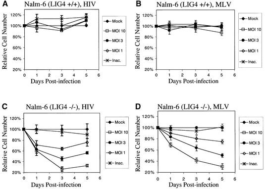
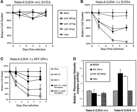
Early after infection, the retroviral RNA genome is reverse transcribed to generate a linear cDNA copy, then that copy is integrated into a chromosome of the host cell. We report that unintegrated viral cDNA is a substrate for the host cell non-homologous DNA end joining (NHEJ) pathway, which normally repairs cellular double-strand breaks by end ligation. NHEJ activity was found to be required for an end-ligation reaction that circularizes a portion of the unintegrated viral cDNA in infected cells. Consistent with this, the NHEJ proteins Ku70 and Ku80 were found to be bound to purified retroviral replication intermediates. Cells defective in NHEJ are known to undergo apoptosis in response to retroviral infection, a response that we show requires reverse transcription to form the cDNA genome but not subsequent integration. We propose that the double-strand ends present in unintegrated cDNA promote apoptosis, as is known to be the case for chromosomal double-strand breaks, and cDNA circularization removes the pro-apoptotic signal.


Figures

None
Fig. 1. Retroviral cDNA forms produced after infection. Retroviral genomes contained in preintegration complexes (PICs) can be integrated to generate an integration intermediate (II) which is then repaired to yield a provirus. Alternatively, the cDNA can be circularized to form 2-LTR or 1-LTR circles.
None
Fig. 2. The NHEJ system is required for formation of 2-LTR circles. (A) Southern blot analysis of extrachromosomal HIV cDNA forms. (B) Southern blot analysis of extrachromosomal MLV cDNA forms. Unintegrated cDNA was isolated from the cell lines indicated above the autoradiograms after high titer infection (m.o.i. 5) using the Hirt procedure (Arad, 1998). Expected mobilities of DNAs from the unintegrated DNA forms are as indicated beside the gel (L-LTR is the left half of the linear genome, R-LTR is the right half). For the HIV-1 vector, the viral cDNA molecules were digested with XhoI and BamHI. For the MLV vector, the viral cDNAs were digested with NotI and ClaI. A 32P-labeled DNA matching each cognate LTR was used as probe.
None
Fig. 3. Ku is associated with PICs. (A) Western blot analysis of proteins in purified MLV PIC fractions. Top: Nycodenz gradient analysis of viral genomes and total protein. Parallel fractions from mock-infected NIH 3T3 cells prepared identically are analyzed in the right lane in each panel. The anti-Ku70 and 80 lanes were probed with antibodies against both proteins. (B) Western blot analysis of proteins in the HIV PIC peak fractions. For the integrase blot the dot indicates HIV-integrase (the upper band is present in the control and represents a contaminating cellular protein). Only Ku80 was analyzed. (C) Immunoprecipitation (IP) of HIV vector PICs. (D) IP of MLV PICs. Lane 1, pre-immune serum; lane 2, anti-Ku80 antibody. PICs captured on protein A–agarose beads were exposed to target DNA, then the cDNA products were analyzed on Southern blots probed with the cognate LTR sequences labeled with 32P. ‘II’ indicates the integration intermediate generated by PIC integration, ‘cDNA’ indicates the unintegrated linear form. (E) Lack of IP of PICs with anti-Ku80 antibody from cells mutant in Ku80. PICs were prepared from CHO-K1 (lanes 1–3) or xrs6 cells (Ku-mutant; lanes 4–6). Load control, initial input PIC fraction; pre-immune, control IP with pre-immune serum; anti-Ku80, IP with anti-Ku80 antibody.
None
Fig. 4. Lack of requirement for Ku80 during integration by PICs in vitro. The cell lines indicated above the autoradiograms were infected with HIV vectors (A) or MLV vectors (B) and PICs isolated. The PICs were mixed in vitro with target DNA (lanes 2, 4 and 6) or not (lanes 1, 3 and 5) and formation of the integration intermediate (labeled ‘II’, see Figure 1) quantitated by PhosphorImager.
None
Fig. 5. Toxicity of retroviral infection in NHEJ-mutant (Nalm-6 LIG4–/–) cells. (A) Infection of Nalm-6 with the HIV-based vector. The m.o.i. of 10 corresponds to 60 ng of p24 capsid protein per 5 × 10⁵ cells. (B) Infection of Nalm-6 with the MLV vector. (C) Infection of Nalm-6 LIG4–/– with the HIV vector. (D) Infection of Nalm-6 LIG4–/– with the MLV vector. Viable cell numbers were normalized to the mock-infected control. For the cytotoxicity experiments, an HIV vector containing the central polypurine tract was used to boost the titer (Follenzi et al., 2000). M.o.i. values reflect titers (gfp-transducing units per ml) on 293T cells. Each point represents the average of three separate cultures.
None
Fig. 6. Apoptosis of NHEJ-mutant cells (Nalm-6 LIG4–/–) after infection requires viral reverse transcription but not integration. (A) Infection of Nalm-6 cells with the HIV (E152A) vector. (B) Infection of Nalm-6 LIG4–/– with the HIV (E152A) vector. (C) Addition of 10 µM nevirapine inhibits the toxic effect of HIV (IN+) infection in Nalm-6 LIG4–/– cells. (D) Assay of caspase 3 activation after infection. Caspase 3 enzyme was captured by an immobilized anti-caspase 3 antibody, then a fluorogenic caspase 3 substrate added. Fluorescence intensity measured after incubation was normalized to the mock-infected control. Infections were initiated by adding 60 ng of p24 capsid antigen per 5 × 10⁵ cells.
None
Fig. 7. Characterization of the integrase-mutant HIV (E152A)-gfp vector. (A) Fields of Nalm-6 cells infected with the HIV-gfp vector or HIV (E152A)-gfp vector analyzed 14 days after infection. Left column, phase contrast image of infected cells. Right column, fluorescent image of the same fields. (B) Quantitative PCR analysis of reverse transcription and integration by HIV (IN+) and HIV (E152A) in Nalm-6 cells. Filled symbols (‘RT’), quantitation of HIV genomes; open symbols (‘provirus’), quantitation of integrated proviruses by Alu-PCR. The approach to quantitating results of Alu-PCR is described in Butler et al. (2001). Circles, HIV (IN+); squares, HIV (E152A). (C) Southern blot analysis of DNA from Nalm-6 cells infected with the HIV-gfp vector or the HIV (E152A)-gfp vector. Unintegrated (Hirt) cDNA (lanes 1 and 2) was isolated 12 h after infection and analyzed by Southern blotting. Genomic DNA (lanes 3 and 4) was isolated 14 days after infection and cleaved with BamHI and EcoRI, which each cut once in the vector DNA. The blots were probed with a 32P-labeled sequence matching the internal vector sequences. Infections were initiated by adding 60 ng of p24 capsid antigen per 5 × 10⁵ cells.

References

    1. Arad U. (1998) Modified Hirt procedure for rapid purification of extrachromosomal DNA from mammalian cells. Biotechniques, 24, 760–762. - PubMed
    1. Baekelandt V., Claeys,A., Cherepanov,P., De Clercq,E., Strooper,B.D., Nuttin,B. and Debyser,Z. (2000) DNA-dependent protein kinase is not required for efficient lentivirus integration. J. Virol., 74, 11278–11285. - PMC - PubMed
    1. Beall E.L. and Rio,D.C. (1996) Drosophila IRBP/Ku p70 corresponds to the mutagen-sensitive mus309 gene and is involved in P-element excision in vivo. Genes Dev., 10, 921–933. - PubMed
    1. Bowerman B., Brown,P.O., Bishop,J.M. and Varmus,H.E. (1989) A nucleoprotein complex mediates the integration of retroviral DNA. Genes Dev., 3, 469–478. - PubMed
    1. Brown P.O., Bowerman,B., Varmus,H.E. and Bishop,J.M. (1987) Correct integration of retroviral DNA in vitro. Cell, 49, 347–356. - PubMed

Publication types

MeSH terms