. 2001 Jun 15;20(12):3282-91.
doi: 10.1093/emboj/20.12.3282.

DNA helicase-mediated packaging of adeno-associated virus type 2 genomes into preformed capsids

Affiliations

DNA helicase-mediated packaging of adeno-associated virus type 2 genomes into preformed capsids

J A King et al. EMBO J. .

Abstract

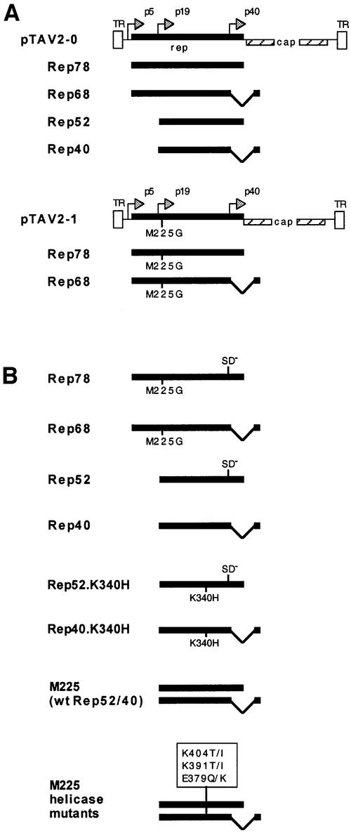
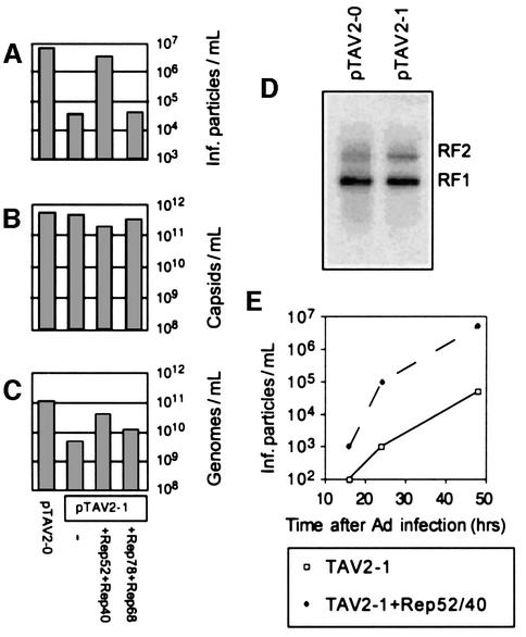
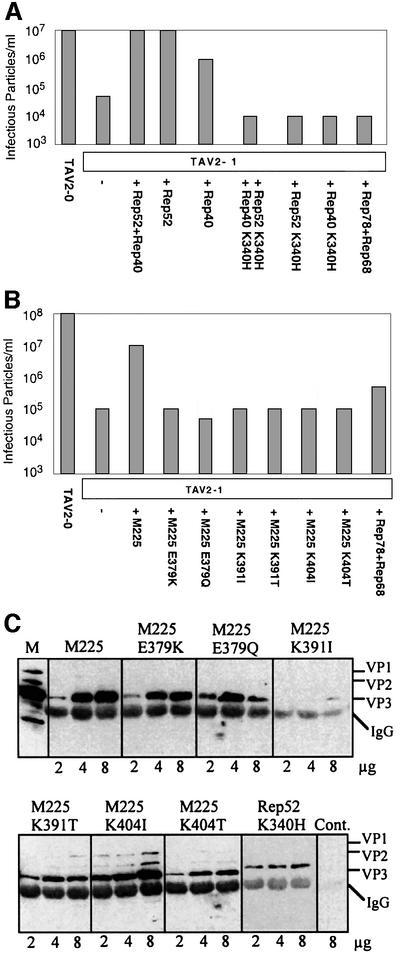
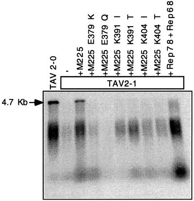
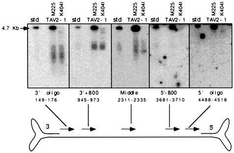
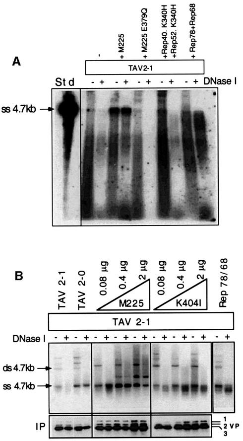
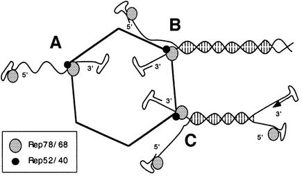
Helicases not only catalyse the disruption of hydrogen bonding between complementary regions of nucleic acids, but also move along nucleic acid strands in a polar fashion. Here we show that the Rep52 and Rep40 proteins of adeno-associated virus type 2 (AAV-2) are required to translocate capsid-associated, single-stranded DNA genomes into preformed empty AAV-2 capsids, and that the DNA helicase function of Rep52/40 is essential for this process. Furthermore, DNase protection experiments suggest that insertion of AAV-2 genomes proceeds from the 3' end, which correlates with the 3'-->5' processivity demonstrated for the Rep52/40 helicase. A model is proposed in which capsid-immobilized helicase complexes act as molecular motors to 'pump' single-stranded DNA across the capsid boundary.


Figures

None
Fig. 1. AAV viral constructs and Rep expression plasmids. (A) pTAV2-0 consists of the entire AAV-2 genome, including both inverted terminal repeats (TR). pTAV2-1 is derived from pTAV2-0, but has the Rep52/40 translation start site methionine 225 mutated to glycine. Spliced and unspliced gene products expressed from the rep open reading frames (ORFs) of the two virus genomes are also shown. AAV promoters are denoted by p5, p19 and p40. (B) The Rep78, Rep68, Rep52 and Rep40 proteins are individually expressed from a CMV promoter within the pKEX-XL plasmid (Hörer et al., 1995). The K340H small Rep mutants contain a lysine to histidine mutation at amino acid 116 (corresponding to amino acids 340 of the complete Rep ORF), which mutates the ATP binding site (Chejanovsky and Carter, 1990). M225 is also based on pKEX-XL and allows expression of both Rep52 and Rep40, as the splice sites have not been altered. A series of helicase mutations (E379Q, E379K, K391I, K391T, K404I, K404T) were introduced into M225 as described in Materials and methods. SD denotes a mutated splice donor site.
None
Fig. 2. Requirement of Rep52/40 for efficient DNA encapsidation. Viral supernatants were generated from cells transfected either with the wt pTAV2-0 construct or with the mutant pTAV2-1 construct and in combination with plasmids expressing either the small or large Rep proteins. Supernatants were assayed for (A) infectious viral titre, (B) ELISA-based AAV-2 capsid titre and (C) quantity of encapsidated AAV-2 DNA. (D) Low molecular weight RF (replication form) DNAs were isolated from cells transfected with pTAV2-0 or pTAV2-1 using a modified Hirt extraction procedure, and detected by Southern blotting using an AAV rep-specific probe. (E) Virus production was measured over time in the presence and absence of Rep52/40.
None
Fig. 3. Effect of small Rep DNA helicase on AAV DNA packaging. (A) Cells were transfected either with pTAV2-0 or with pTAV2-1, either alone or together with plasmids encoding wt or small Rep proteins mutated in the helicase domain (K340H) or large Rep proteins. Extracts were prepared and their infectious particle titres determined. (B) A similar analysis was carried out using plasmids expressing either the wt or mutant forms of both small Rep proteins, in which point mutations had been introduced at additional conserved amino acid residues of the helicase domain (see Figure 1). (C) The capacity of these mutants to form complexes with AAV-2 capsid proteins was tested by co-transfecting increasing amounts (2, 4 and 8 µg) of the capsid protein-expressing plasmid (CMV-VP) with a fixed amount (3 μg) of each small Rep expression plasmid. Complexes were immunoprecipitated from cell extracts using an anti-Rep antiserum and analysed via western blotting using a monoclonal anti-VP antibody to detect capsid proteins. A control reaction was carried out using extracts of cells that had not been transfected with a Rep-expressing plasmid (Cont). M, standard showing the expression of the three capsid proteins (VP1–3). IgG indicates the position of the immunoglobulin heavy chain of the antiserum used for the immunoprecipitation.
None
Fig. 4. Genome encapsidation in the presence of Rep52/40 DNA helicase mutants. Viral genomes from virus preparations (see Figure 3B) were isolated from the virus supernatants after extensive DNase I digestion, separated by alkaline agarose gel electrophoresis and Southern blotted. AAV DNA was detected using a rep-specific probe. 4.7 kb indicates full-length encapsidated AAV DNA.
None
Fig. 5. Directionality of AAV packaging. Cells were transfected with the small Rep-deleted pTAV2-1 plasmid and either the wt Rep52/40 (M225) or the K404I helicase mutant Rep52/40 construct. DNase I-protected genomes from lysates were separated on alkaline agarose gels. Following Southern blotting, partially and fully packaged genomes were detected using end-labelled oligonucleotides that hybridize to the 3′ end (3′ oligo), a region 800 bp from the 3′ end (3′+800), a central region (middle), a region 800 bp from the 5′ end (5′–800) and the 5′ end (5′ oligo) of one of the two packagable genome strands, as indicated (arrowheads point in the 5′→3′ direction).
None
Fig. 6. Analysis of capsid-associated and encapsidated genomes. (A) Viral supernatants obtained from cells that had been transfected with pTAV2-1 alone, or together with either both large Rep expression plasmids (Rep78/68), wt (M225) or helicase mutant (E379Q or K340H) small Rep expression plasmids, were digested (+) or not (–) with DNase I to discriminate between capsid-associated and packaged genomes. Capsids were then immunoprecipitated from each extract using equal amounts of protein A–Sepharose-bound A20 antibody. Coprecipitated genomes were isolated and electrophoresed on an alkaline agarose gel, Southern blotted and detected using a rep-specific probe. A 4.7 kb AAV genome fragment was used as a standard (Std). (B) Cells were transfected with pTAV2-0 or pTAV2-1 alone or with pTAV2-1 and both large Rep expression plasmids (Rep78/68) or increasing amounts of either the wt Rep52/40 (M225) plasmid or the M225-K404I helicase mutant. Cell extracts were then processed as described in (A). Viral genomes were extracted and electrophoresed on neutral agarose gels, Southern blotted and detected using a random-primed, rep-specific probe. A portion of each immunoprecipitate was tested for the level of capsid recovery via western analysis.
None
Fig. 7. Model for the involvement of Rep helicases as genome packaging motors. Most helicases bind to ssDNA adjacent to duplex regions and then proceed to unwind the two strands, moving along the bound single strand, under the consumption of ATP. The AAV packaging complex represents an immobilized helicase complex, composed of large and small Rep proteins, on the capsid surface. Both ss and ds genomes are able to bind to the packaging complex via interaction with the large Rep proteins, which bind sequence specifically to both ends and covalently to the 5′ end. The genome is then translocated through the packaging complex and into the capsid either (A) as a single-stranded molecule using the initial ‘scanning’ function before the first duplexed base pairs are encountered or (B) by unwinding a ds dimer or multimer genome on the capsid surface at the same time. (C) Simultaneous replication (arrow) of a ds monomer genome being packaged would result in premature strand displacement. Large Rep molecules at 5′ ends are covalently bound.

References

    1. Baker T.A. and Bell,S.P. (1998) Polymerases and the replisome: machines within machines. Cell, 92, 295–305. - PubMed
    1. Berns K.I. (1990) Parvovirus replication. Microbiol. Rev., 54, 316–329. - PMC - PubMed
    1. Bina M., Ng,S.C. and Blasquez,V. (1983) Simian virus 40 chromatin interaction with the capsid proteins. J. Biomol. Struct. Dyn., 1, 689–704. - PubMed
    1. Bird L.E., Subramanya,H.S. and Wigley,D.B. (1998) Helicases: a unifying structural theme? Curr. Opin. Struct. Biol., 8, 14–18. - PubMed
    1. Black L.W. (1989) DNA packaging in dsDNA bacteriophages. Annu. Rev. Microbiol., 43, 267–292. - PubMed

Publication types