. 2001 Jun;12(6):1611-21.
doi: 10.1091/mbc.12.6.1611.

SPO21 is required for meiosis-specific modification of the spindle pole body in yeast

Affiliations
Free PMC article

SPO21 is required for meiosis-specific modification of the spindle pole body in yeast

B K Bajgier et al. Mol Biol Cell. 2001 Jun.
Free PMC article

Abstract

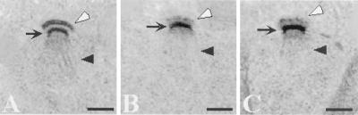
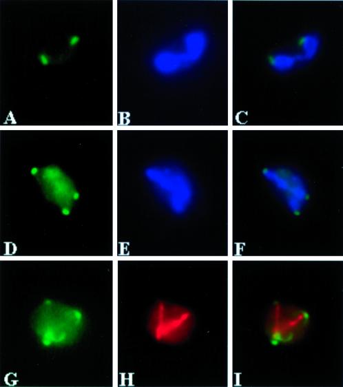
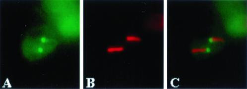
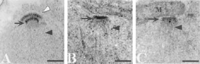
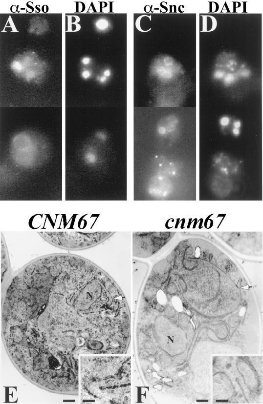
During meiosis II in the yeast Saccharomyces cerevisiae, the cytoplasmic face of the spindle pole body changes from a site of microtubule initiation to a site of de novo membrane formation. These membranes are required to package the haploid meiotic products into spores. This functional change in the spindle pole body involves the expansion and modification of its cytoplasmic face, termed the outer plaque. We report here that SPO21 is required for this modification. The Spo21 protein localizes to the spindle pole in meiotic cells. In the absence of SPO21 the structure of the outer plaque is abnormal, and prospore membranes do not form. Further, decreased dosage of SPO21 leaves only two of the four spindle pole bodies competent to generate membranes. Mutation of CNM67, encoding a known component of the mitotic outer plaque, also results in a meiotic outer plaque defect but does not block membrane formation, suggesting that Spo21p may play a direct role in initiating membrane formation.


Figures

Figure 1
Figure 1
Alignment of Spo21p and Spc72p sequences. The alignment was prepared with the use of the BLAST 2 algorithm (Tatusova and Madden, 1999). The residues at the amino terminal ends of the predicted coiled-coil regions, Arg283 of Spo21p and His359 of Spc72p, are highlighted.
Figure 2
Figure 2
spo21 mutants show no defect in meiotic progression. Strains AN120 and AN180 were transferred to 2% KOAc, and at indicated times samples were removed and fixed, and progression through meiosis was examined by DAPI staining. ×, AN120, mononucleate cells; ♦, AN180 mononucleate cells; ▪, AN120 binucleate and tetranucleate cells; ∗, AN180 binucleate and tetranucleate cells; ▴, AN180 cells displaying nuclear fragmentation.
Figure 3
Figure 3
No prospore membranes are formed in a spo21Δ mutant. Strains AN120 (SPO21/SPO21) and AN180 (spo21Δ/spo21Δ) were transferred to 2% KOAc and analyzed by indirect immunofluorescence as described in MATERIALS AND METHODS. (A, E, and I) AN120 stained with anti-Ssop, anti-Sncp, and anti-Spr3p antibodies, respectively. (B, F, and J) DAPI staining of the corresponding cells in A, E, and I. (C, G, and K) AN180 stained with anti-Ssop, anti-Sncp, and anti-Spr3p antibodies, respectively. (D, H, and L) DAPI staining of the corresponding cells in C, G, and K, demonstrating that the cells analyzed have undergone meiosis II. Images in E, F, G, H, K, and L are composites.
Figure 4
Figure 4
Prospore membranes form on opposite spindles in a spo21::GFP strain. Strain AN230 (spo21::GFP/spo21::GFP) was transferred to 2% KOAc and analyzed by fluorescence microscopy as described in MATERIALS AND METHODS. (A) Anti-Ssop staining. (B) Antitubulin staining of the cells in A. (C) DAPI staining of the same cells.
Figure 5
Figure 5
SPO21 is required for proper modification of the outer plaque. Strains AN120 (SPO21/SPO21) and AN180 (spo21Δ/spo21Δ) were transferred to 2% KOAc and prepared for transmission EM as described in MATERIALS AND METHODS. (A) Wild-type meiosis II SPB. (B and C) Meiosis II SPBs in the spo21 mutant. Clear arrowhead, outer plaque; arrow, central plaque; dark arrowhead, spindle microtubules. Scale bar, 200 nm.
Figure 6
Figure 6
Spo21-GFP localizes to the spindle pole. Strain AN230 (spo21::GFP/spo21::GFP) carrying pRS316-SPO21::GFP2 was transferred to 2% KOAc and analyzed by fluorescence microscopy as described in MATERIALS AND METHODS. (A) Spo21-GFP in a meiosis I cell. (B) DAPI staining of the same cell in A. (C) Merged image of A and B showing localization of Spo21-GFP to the periphery of the nucleus. (D) Spo21-GFP foci in a meiosis II cell. (E) DAPI staining of the same cell in D. (F) Merged image of D and E showing Spo21-GFP near the leading edge of the segregating chromatin. (G) A meiosis II cell showing four Spo21-GFP foci. (H) Antitubulin staining of the same cell in G. (I) Merged image of G and H, demonstrating the Spo21-GFP foci are at the spindle poles.
Figure 7
Figure 7
Spo21p is found on the poles of opposite spindles in during NSD formation. Strain AN230 (spo21:: GFP/spo21::GFP) carrying pRS316-SPO21::GFP2 was subjected to interrupted sporulation and analyzed by fluorescence microscopy as described in MATERIALS AND METHODS. (A) Localization of Spo21-GFP. (B) Antitubulin staining of the cell in A. (C) Merged image of A and B showing localization of Spo21-GFP to the poles of opposite spindles.
Figure 8
Figure 8
Outer plaques are absent in the cnm67 mutant. Strains AN120 (SPO21/SPO21) and AN161 (cnm67Δ/cnm67Δ) were transferred to 2% KOAc and prepared for transmission EM as described in MATERIALS AND METHODS. (A) Wild-type meiosis II SPB. (B and C) Meiosis II SPBs in the cnm67 mutant. Clear arrowhead, outer plaque; arrow, central plaque; dark arrowhead, spindle microtubules. M, mitochondrion; Scale bar, 200 nm.
Figure 9
Figure 9
Prospore membranes form in abnormal locations in a cnm67 mutant. Strain AN161 (cnm67Δ/cnm67Δ) was transferred to 2% KOAc and prepared for immunofluorescence and transmission EM as described in MATERIALS AND METHODS. (A) Anti-Ssop staining. (B) DAPI staining of cells in A. (C) Anti-Sncp staining. (D) DAPI staining of cells in C. (A–D) Composite images. (E) Transmission EM of a wild-type cell prepared by permanganate staining shows a prospore membrane engulfing a nuclear lobe (arrowhead). (F) A cnm67 mutant prepared similarly to the cell in E displays a prospore membrane (arrowhead) unassociated with nucleus (N). Insets: Higher magnification showing the characteristic knob on the end of the prospore membranes in E and F. Scale bars: large panels, 500 nm; insets, 150 nm.

References

    1. Adams IR, Kilmartin JV. Localization of core spindle pole body (SPB) components during SPB duplication in Saccharomyces cerevisiae. J Cell Biol. 1999;145:809–823. - PMC - PubMed
    1. Arvan P, Castle D. Sorting and storage during secretory granule biogenesis: looking backward and looking forward. Biochem J. 1998;332:593–610. - PMC - PubMed
    1. Brachat A, Kilmartin JV, Wach A, Philippsen P. Saccharomyces cerevisiae cells with defective spindle pole body outer plaques accomplish nuclear migration via half-bridge-organized microtubules. Mol Biol Cell. 1998;9:977–991. - PMC - PubMed
    1. Burgoyne RD, Morgan A. Regulated exocytosis. Biochem J. 1993;293:305–316. - PMC - PubMed
    1. Byers B. Cytology of the yeast life cycle. In: Strathern JN, Jones EW, Broach JR, editors. The Molecular Biology of the Yeast Saccharomyces: Life Cycle and Inheritance. Cold Spring Harbor, NY: Cold Spring Harbor Press; 1981. pp. 59–96.

Publication types

MeSH terms