. 2001 Jun;12(6):1623-31.
doi: 10.1091/mbc.12.6.1623.

Lysosomal hydrolase mannose 6-phosphate uncovering enzyme resides in the trans-Golgi network

Affiliations
Free PMC article

Lysosomal hydrolase mannose 6-phosphate uncovering enzyme resides in the trans-Golgi network

J Rohrer et al. Mol Biol Cell. 2001 Jun.
Free PMC article

Abstract

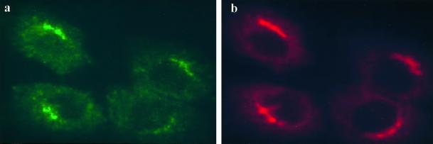
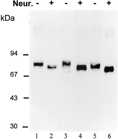
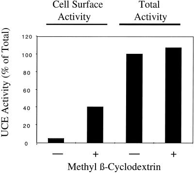
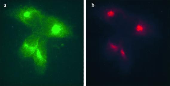
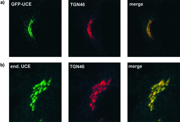
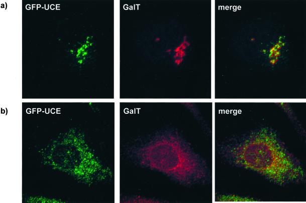
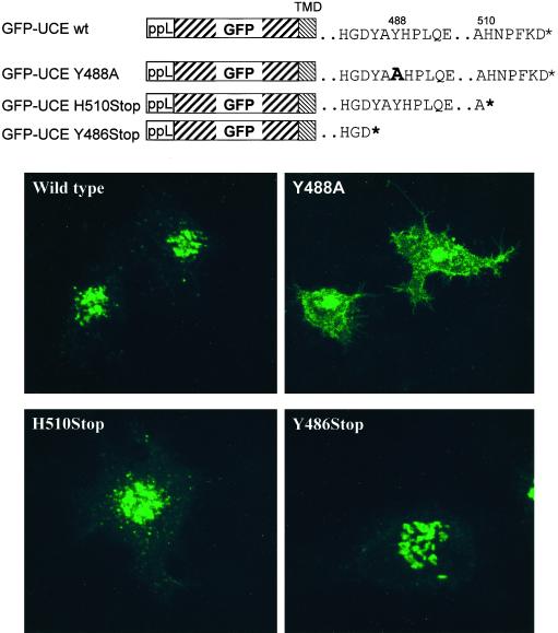
A crucial step in lysosomal biogenesis is catalyzed by "uncovering" enzyme (UCE), which removes a covering N-acetylglucosamine from the mannose 6-phosphate (Man-6-P) recognition marker on lysosomal hydrolases. This study shows that UCE resides in the trans-Golgi network (TGN) and cycles between the TGN and plasma membrane. The cytosolic domain of UCE contains two potential endocytosis motifs: (488)YHPL and C-terminal (511)NPFKD. YHPL is shown to be the more potent of the two in retrieval of UCE from the plasma membrane. A green-fluorescent protein-UCE transmembrane-cytosolic domain fusion protein colocalizes with TGN 46, as does endogenous UCE in HeLa cells, showing that the transmembrane and cytosolic domains determine intracellular location. These data imply that the Man-6-P recognition marker is formed in the TGN, the compartment where Man-6-P receptors bind cargo and are packaged into clathrin-coated vesicles.


Figures

Figure 1
Figure 1
UCE colocalizes with TGN38 in MDBK cells. Cells were fixed and permeabilized and double labeled with mAb UC-1 to bovine UCE (a, green) and rabbit antibody to TGN38 (b, red).
Figure 2
Figure 2
The human UCE expressed by stably transfected L-cells is sialylated. Membrane extracts of cells expressing human UCE wild type (lanes 1 and 2), Y488A mutant (lanes 3 and 4), or H510 stop mutant (lanes 5 and 6) were treated (+; lanes 2, 4, and 6) or not treated (−; lanes 1, 3, and 5) with Vibrio cholera neuraminidase (Neur.) for 80 min at 37°C. The reaction mixtures were boiled in SDS-PAGE sample buffer and subjected to reducing SDS-PAGE on a 7.5% gel. The gel was blotted to nitrocellulose, which was probed with affinity-purified rabbit anti-human UCE antibody (1:1000) and detected by enhanced chemiluminescence.
Figure 3
Figure 3
The effect of methyl β-cyclodextrin on surface expression of UCE. L-cells expressing wild-type human UCE were treated (+) or not (−) with 10 mM methyl β-cyclodextrin for 2 h before assaying for UCE activity with or without methyl β-cyclodextrin, and with Triton X-100 (total) or without Triton X-100 (cell surface) as described in MATERIALS AND METHODS. The activity of UCE is expressed as the percentage of total activity in the non-methyl β-cyclodextrin treated and is the average of two separate experiments.
Figure 4
Figure 4
UCE cycles from the plasma membrane to the TGN. MDBK cells were incubated with antibody UC-1 for 2 h at 37°C, fixed, permeabilized, and probed with anti-mouse antibody (a, green) and also double labeled with rabbit antibody to TGN38 (b, red).
Figure 5
Figure 5
GFP-UCE and endogenous UCE both colocalize with TGN 46. (a) HeLa cells stably transfected with GFP-UCE-wild-type TM-cytosolic tail were fixed, permeabilized, and labeled with sheep anti-human TGN 46, followed by goat anti-sheep Alexa fluor 594. Left, GFP-UCE; middle, TGN 46; right, merged image. (b) Nontransfected HeLa cells were double labeled with rabbit anti-human UCE and sheep anti-human TGN 46, followed by goat anti-rabbit Alexa 488 and donkey anti-sheep Alexa 594, respectively. Left, UCE; middle, TGN 46; right, merged image.
Figure 6
Figure 6
GFP-UCE does not colocalize with galactosyltransferase (GalT). HeLa cells stably transfected with GFP-UCE wild type were labeled with anti-galactosyltransferase antibody after incubation for 30 min in the absence of BFA (a) or after 30 min of incubation with BFA (b). Left, GFP-UCE; middle, galactosyltransferase; right, merged image.
Figure 7
Figure 7
The Y488A mutation in GFP-UCE inhibits its endocytosis from the plasma membrane. HeLa cells were transiently transfected with GFP-UCE TM-cytosolic tail constructs: wild type (wt), Y488A, H510Stop, or Y486Stop. The diagrams show the structures of these constructs: ppL is the preprolactin signal sequence followed by GFP, the transmembrane domain (TMD) of UCE, and the cytosolic tail of UCE as modified in the various mutants. The * indicates the end of the cytosolic tail.

Similar articles

Cited by

References

    1. Benmerah A, Begue B, Dautry-Varsat A, Cerf-Bensussan N. The ear of α-adaptin interacts with the COOH-terminal domain of the Eps 15 protein. J Biol Chem. 1996;271:12111–12116. - PubMed
    1. Berger EG, Aegerter E, Mandel T, Hauri HP. Monoclonal antibodies to soluble, human milk galactosyltransferase (lactose synthase A protein) Carbohydr Res. 1986;149:23–33. - PubMed
    1. Chapman RE, Munro S. Retrieval of TGN proteins from the cell surface requires endosomal acidification. EMBO J. 1994;13:2305–2312. - PMC - PubMed
    1. Colley KJ. Golgi localization of glycosyltransferases: more questions than answers. Glycobiology. 1997;7:1–13. - PMC - PubMed
    1. Deutscher SL, Creek KE, Merion M, Hirschberg CB. Subractionation of rat liver Golgi apparatus: separation of enzyme activities involved in the biosynthesis of the phosphomannosyl recognition marker in lysosomal enzymes. Proc Natl Acad Sci USA. 1983;80:3938–3942. - PMC - PubMed

Publication types

MeSH terms

Substances

LinkOut - more resources