. 2001 Jun;12(6):1645-69.
doi: 10.1091/mbc.12.6.1645.

Dynamic localization of the Swe1 regulator Hsl7 during the Saccharomyces cerevisiae cell cycle

Affiliations
Free PMC article

Dynamic localization of the Swe1 regulator Hsl7 during the Saccharomyces cerevisiae cell cycle

V J Cid et al. Mol Biol Cell. 2001 Jun.
Free PMC article

Abstract

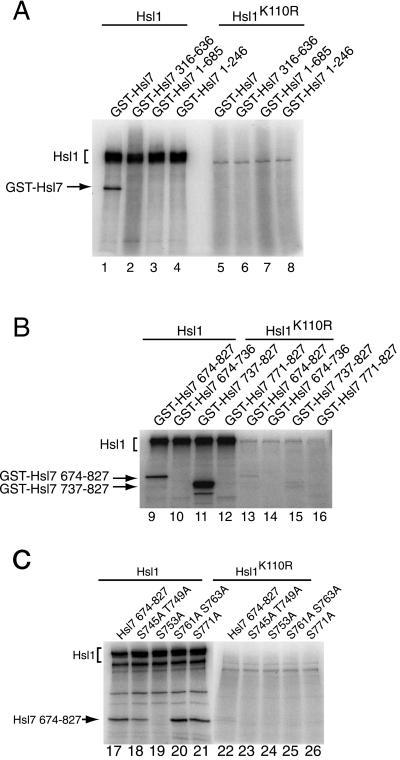
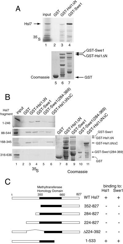
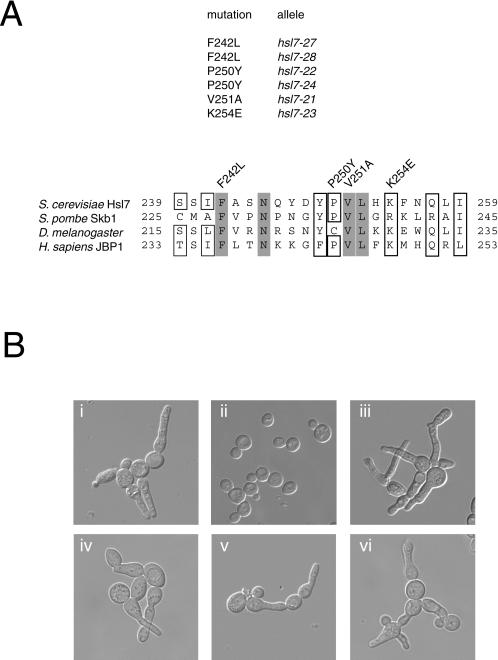
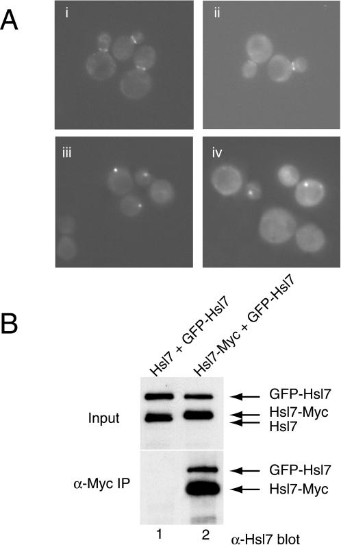
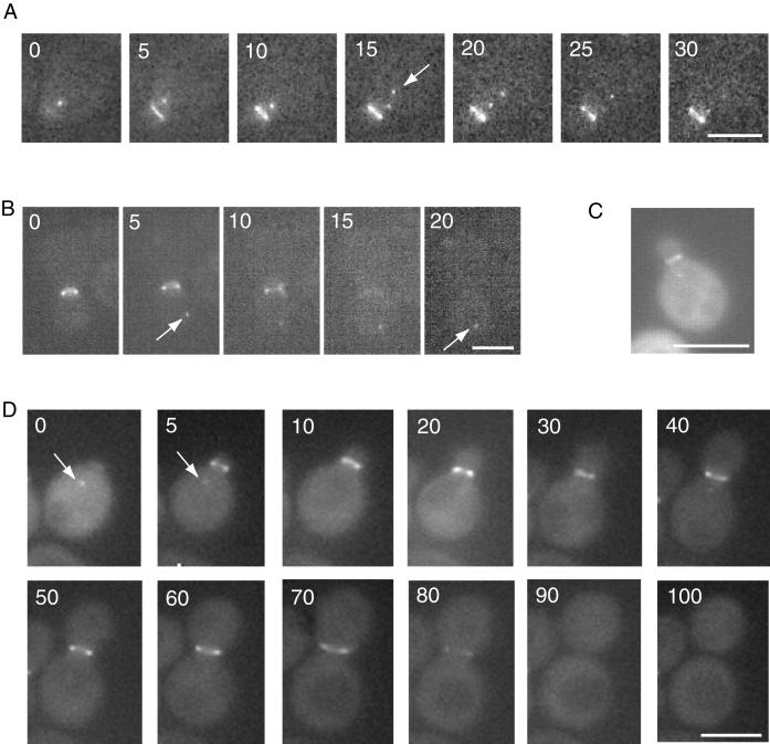
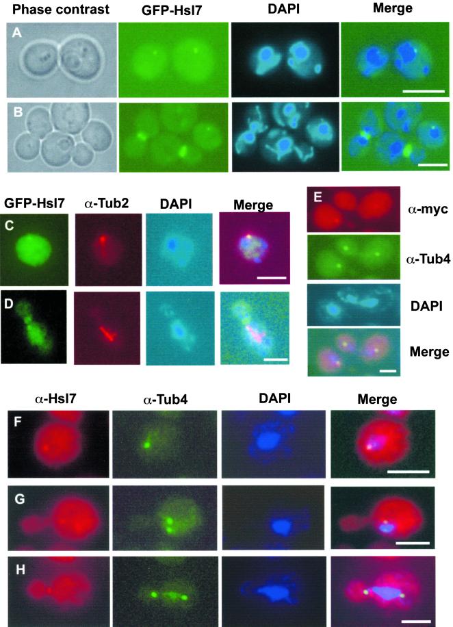
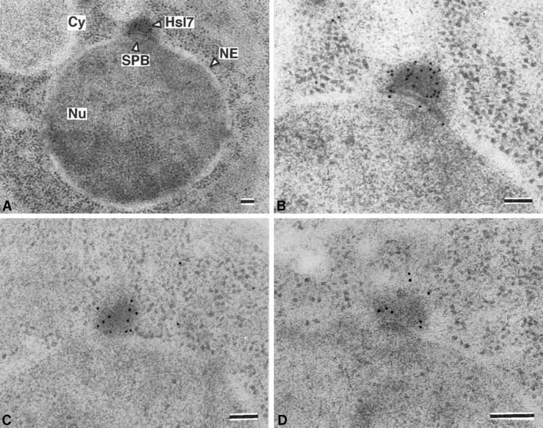
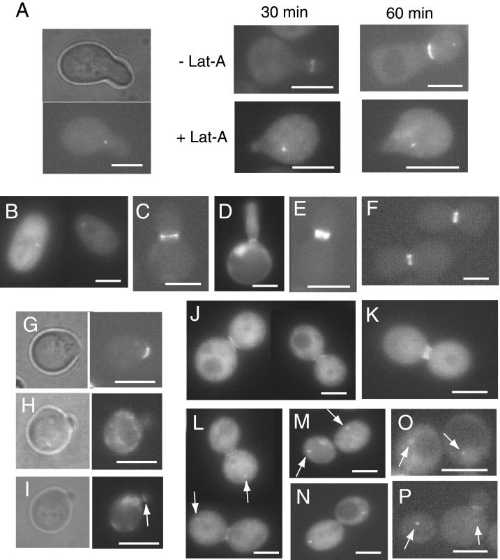
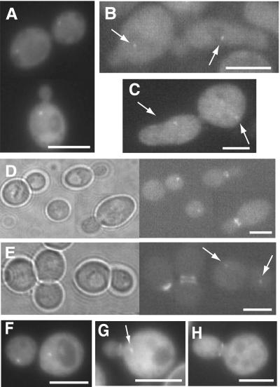
In Saccharomyces cerevisiae, entry into mitosis requires activation of the cyclin-dependent kinase Cdc28 in its cyclin B (Clb)-associated form. Clb-bound Cdc28 is susceptible to inhibitory tyrosine phosphorylation by Swe1 protein kinase. Swe1 is itself negatively regulated by Hsl1, a Nim1-related protein kinase, and by Hsl7, a presumptive protein-arginine methyltransferase. In vivo all three proteins localize to the bud neck in a septin-dependent manner, consistent with our previous proposal that formation of Hsl1-Hsl7-Swe1 complexes constitutes a checkpoint that monitors septin assembly. We show here that Hsl7 is phosphorylated by Hsl1 in immune-complex kinase assays and can physically associate in vitro with either Hsl1 or Swe1 in the absence of any other yeast proteins. With the use of both the two-hybrid method and in vitro binding assays, we found that Hsl7 contains distinct binding sites for Hsl1 and Swe1. A differential interaction trap approach was used to isolate four single-site substitution mutations in Hsl7, which cluster within a discrete region of its N-terminal domain, that are specifically defective in binding Hsl1. When expressed in hsl7Delta cells, each of these Hsl7 point mutants is unable to localize at the bud neck and cannot mediate down-regulation of Swe1, but retains other functions of Hsl7, including oligomerization and association with Swe1. GFP-fusions of these Hsl1-binding defective Hsl7 proteins localize as a bright perinuclear dot, but never localize to the bud neck; likewise, in hsl1Delta cells, a GFP-fusion to wild-type Hsl7 or native Hsl7 localizes to this dot. Cell synchronization studies showed that, normally, Hsl7 localizes to the dot, but only in cells in the G1 phase of the cell cycle. Immunofluorescence analysis and immunoelectron microscopy established that the dot corresponds to the outer plaque of the spindle pole body (SPB). These data demonstrate that association between Hsl1 and Hsl7 at the bud neck is required to alleviate Swe1-imposed G2-M delay. Hsl7 localization at the SPB during G1 may play some additional role in fine-tuning the coordination between nuclear and cortical events before mitosis.


Figures

Figure 1
Figure 1
The C terminus of Hsl7 is phosphorylated by Hsl1 protein kinase. Equivalent amounts of protein (1 mg of total) from extracts of a protease-deficient strain (MJY153) expressing either triple-HA-tagged wild-type Hsl1 or a catalytically defective derivative, Hsl1(K110R), from plasmids (YCpLG-HSL1(HA)3 and YCpLG-HLS1-K110R(HA)3, respectively), were subjected to immunoprecipitation with mouse anti-HA mAb HA.11. The resulting immune complexes were resuspended in protein kinase assay buffer, incubated for 10 min at 30°C with Mg2+, [γ-32P]ATP, and a GST-Hsl7 fusion or the indicated C-terminal truncations (purified from yeast) (A), or the indicated C-terminal fragments (purified from bacteria) (B), or the indicated site-directed mutants (purified from bacteria) (C), and then analyzed by SDS-PAGE followed by autoradiography.
Figure 2
Figure 2
Hsl1- and Swe1-binding sites reside in the N-terminal domain of Hsl7. (A) Radiolabeled Hsl7 (35S), prepared by in vitro translation, was incubated with equivalent amounts of glutathione-agarose beads preadsorbed with equivalents amounts of bacterially expressed GST (lane 2), GST-Hsl1(833–1518) (GST-Hsl1ΔN) (lane 3), or GST-Swe1 (lane 4). After washing, the beads were eluted with SDS-PAGE sample buffer and the bound protein analyzed by SDS-PAGE and autoradiography, along with a portion of the 35S-labeled Hsl7 (10% of the amount added in the binding reactions; “input”) (lane 1). Equivalent samples of the beads containing each bacterially produced protein (GST, lane 5; GST-Hsl1(833–1518), lane 6; GST-Swe1, lane 7) were analyzed by SDS-PAGE and staining with Coomassie brilliant blue to demonstrate equal loading. (B) Various fragments of Hsl7 indicated (1–246, 88–544, 168–345, 316–636) were each prepared in radiolabeled form by in vitro translation and incubated, as described in A, with beads carrying GST (lane 2), GST-Hsl1(833–1518) (lane 3), GST-Swe1 (lane 4), GST-Swe1(284–369) (lane 5), and GST-Hsl1(1018–1244; 1482–1518) (GST-Hsl1ΔNΔC). After washing, the beads were eluted with excess glutathione, and the released protein was analyzed by SDS-PAGE and autoradiography, as in A (left panels). Equivalent samples of the beads containing each bacterially produced protein, as indicated, were analyzed by SDS-PAGE and staining with Coomassie brilliant blue to demonstrate equal loading. (C) A ura3 reporter strain (YD116) carrying a GAL1 promoter-dependent URA3 gene was cotransformed with plasmids expressing either Gal4(TAD)-Hsl1(987–1518) or Gal4(TAD)-Swe1(295–819) (Shulewitz et al., 1999) and plasmids expressing Gal4(DBD) fused to the indicated portions of Hsl7. Methyltransferase homology domain denotes the segment of Hsl7 homologous to known protein-arginine methyltransferases (Pollack et al., 1999; Ma, 2000). The ability (+) or inability (−) of each pair of fusions to activate transcription of the GAL1-dependent URA3 reporter gene, as judged by growth in the absence of uracil, is summarized in the right-hand columns.
Figure 3
Figure 3
Point mutations in Hsl7 define its Hsl1-binding site and prevent function. (A) Positions of the different single-residue substitution mutations (and their corresponding allele numbers) that prevent association of Hsl7 with Hsl1, as assessed by the two-hybrid method, are indicated within the primary structure of Hsl7 (and several of its homologues from the organisms indicated). Gray boxes indicate absolutely conserved residues; open boxes indicated highly conserved residues. (B) An hsl7Δ strain (MJY102) was transformed with an empty vector (i) or the same plasmid expressing either normal Hsl7 (ii), Hsl7(F242L) (iii), Hsl7(P250Y) (iv), Hsl7(V251A) (v), or Hsl7(K254E) (vi), grown in liquid medium to midexponential phase, and photographed with the use of differential interference contrast microscopy.
Figure 4
Figure 4
Differential subcellular localization of Hsl7 mutants defective for binding to Hsl1. (A) Two of the Hsl1-binding-defective Hsl7 mutants, Hsl7(P250Y) (i and iii) and Hsl7(F242L) (ii and iv), expressed as GFP fusions from the HSL7 promoter on low copy number TRP1-marked (CEN) plasmids, were introduced into either a wild-type HSL7+ strain (MJY112) (i and ii) or an hsl7Δ strain (MJY151) (iii and iv). The transformants were grown to mid-exponential phase at 30°Cin SCGlc-Trp, and samples of each culture were viewed directly in a fluorescence microscope. (B) Protease-deficient strain (BJ2168) was transformed with plasmids expressing either untagged Hsl7 or a c-Myc-epitope tagged derivative, as indicated, and also cotransformed with a plasmid expressing GFP-Hsl7. Equivalent amounts of protein (1 mg of total) from extracts of these cells (input) were subjected to immunoprecipitation with mouse anti-Myc mAb 9E10, as described in MATERIALS AND METHODS. The resulting immunoprecipitates (α-Myc IP) were resolved by SDS-PAGE and analyzed by immunoblotting with mouse polyclonal anti-Hsl7 (bottom), along with samples (respresenting ∼1% of the amount of extract protein that was subjected to immunoprecipitation) (top).
Figure 5
Figure 5
Dynamic relocalization of Hsl7 during the cell division cycle. Time-lapse fluorescence microscopy of live hsl7Δ cells (MJY102) harboring YCpT-GFP-Hsl7(1–685) during bud emergence and early stages of the cell cycle (A) or during cytokinesis (B). Arrows indicate appearance of a cytoplasmic dot. (C) Wild-type strain (MJY112) harboring YCpLG-GFP-HSL7 was grown on SCRaf-Leu medium at 30°C to mid-exponential phase and then galactose was added (2% final concentration). After 5 h of induction, the live cells were viewed in the fluorescence microscope. (D) Time-lapse fluorescence mincroscopy of a live wild-type cell (MJY112) expressing GFP-Hsl7 (full length) from the HSL7 promoter on a CEN plasmid (YCpT-GFP-HSL7) throughout an entire cell cycle, showing cytoplasmic dot (arrow) of GFP-Hsl7 in an unbudded cell whose disappearance and redeposition at the bud neck is concomitant with bud emergence.
Figure 6
Figure 6
Cytoplasmic Hsl7 dot corresponds to the SPB. (A and B) Perinuclear localization of the cytoplasmic dot in G1 cells. Fluorescence microscopy was performed on wild-type cells (MJY112) expressing GFP-Hsl7 (green) from the HSL7 promoter on a CEN plasmid (YCpT-GFP-HSL7) that were grown to mid-exponential phase in SCGlc-Trp at 30°C and stained with a DNA-specific dye (DAPI) to discern the nucleus (blue). (C and D) Wild-type cells (MJY112) expressing GFP-Hsl7 from the GAL1 promoter on a CEN plasmid (YCpLG-GFP-HSL7) were grown in SCRaf-Leu at 30°C to mid-exponential phase and then induced by addition of galactose (2% final concentration). After 6 h, the cells were lightly fixed, permeabilized, stained with rat anti-α-tubulin mAb YOL134 (and an appropriate Cy3-labeled secondary antibody), counterstained with DAPI, and viewed in a fluorescence microscope, as described in MATERIALS AND METHODS. Diffuse greencytoplasmic and nuclear signal is nonspecific autofluorescence, whereas in a G1 cell the prominent green cytoplasmic dot (GFP-Hsl7) is always coincident with a bundle of astral microtubules (C), and in a preanaphase cell, GFP-Hsl7 is always at the bud neck, but can be found occasionally decorating one end of the spindle (D). (E) Wild-type cells (MJY112) expressing C-terminally c-Myc-tagged Hsl7 (Hsl7-Myc) from the GAL1 promoter on a CEN plasmid (YCpUG-HSL7-Myc) were grown in SCRaf-Ura medium at 30°C to mid-exponential phase and then induced with galactose (2% final concentration). After 6 h, cells were fixed, permeabilized, stained with mouse anti-c-Myc mAb 9E10 (visualized with an appropriate Cy3-conjugated secondary antibody) and with rabbit anti-Tub4 polyclonal antibodies (visualized with an appropriate fluorescein isothiocyanate-conjugated secondary antibody), counterstained with DAPI, and visualized in a fluorescence microscope. The cytoplasmic dot of Hsl7-myc is congruent with the γ-tubulin signal in the early G1 cell (lower left) and, in a cell at late stage of mitosis, Hsl7-myc is deposited at one of the developing SPBs (upper right). (F–H) Same cells as in E, except that Hsl7 (red) was stained with affinity-purified mouse polyclonal anti-Hsl7 antibodies (rather than with mouse anti-c-Myc mAb) before costaining with anti-Tub4 antibodies (green) and counterstaining with DAPI. Unbudded (G1) cells always show a single dot of Hsl7 congruent with Tub4 (F), whereas, characteristically, early after SPB duplication and separation Hsl7 associates assymetrically with only one SPB (G). By the time a cell has initiated mitosis, Hsl7 is found exclusively at the bud neck and is never observed at either SPB (H). Bars, 5 μm.
Figure 7
Figure 7
Hsl7 is specifically localized to the cytosolic face of the SPB. Strain MJY112 harboring YCpUG-HSL7-Myc was induced with galactose for 5 h and then prepared for immunoelectron microscopy, as described in MATERIALS AND METHODS. Frozen thin-sections were stained with affinity-purified mouse polyclonal anti-Hsl7 antibodies then with gold particles coated with donkey anti-mouse immunoglobulin, and examined by transmission electron microscopy. In sections of G1 cells, staining was specific for the SPB and no detectable staining of any other place or structure was observed (A). Staining of the SPB was reproducibly observed in every G1 cell examined (B–D). Cy, cytoplasm; NE, nuclear envelope; Nu, nucleus. Bars, 100 nm.
Figure 8
Figure 8
Cell-cycle stage dependence and functional requirements for relocalization of Hsl7. (A) Hsl7 resides at the SPB in cells arrested in G1 by mating pheromone treatment. Strain MJY155 harboring YCpT-GFP-HSL7, expressing GFP-Hsl7 from the HSL7 promoter on a CEN plasmid, was synchronized by exposure to α-factor and viewed in the fluoresence microscope to confirm formation of the characteristic “shmoo” morphology (left). The arrested cells were then washed to release them from the pheromone-imposed block and allowed to resume growth for the indicated times in the absence (−) or presence (+) of latrunculin-A (right). (B) Strain CWY78 (cdc28–4ts) harboring YCpLG-GFP-HSL7 was induced with galactose and shifted to restrictive temperature (37°C) for 4 h. (C) Strain Y543 (cdc4–1ts) harboring YCpT-GFP-HSL7 was shifted to 37°C for 3 h. (D) Strain YSS19 (cdc34–1ts) harboring YCpLG-GFP-HSL7 was induced with galactose and shifted to 37°C for 9 h. (E) Strain VBY610 (cdc4–1 hsl7Δ) harboring YCpT-GFP-Hsl7(1–685) and shifted to 37°C for 3 h. Note that, although this C-terminally truncated version of Hsl7 can be found at one or both SPBs during S phase (as well as at the bud neck) (Figure 5A), Hsl7(1–685) nonetheless relocates quantitatively to the septin rings in cdc4–1 cells arrested at restrictive temperature in which initiation of DNA replication is blocked. (F) Strain VBY3113a (cdc31–1ts hsl7Δ) harboring YCpT-GFP-Hsl7(1–685) and shifted to 37°C for 3 h. (G) Strain HVY21 (rho1–104ts) expressing Cdc10-GFP from plasmid pLA10 (Cid et al., 1998) shifted to 37°C for 90 min. (H) Same strain as in G harboring YCpT-HSL1-GFP under identical conditions; in most cells (91%), like the one shown here, Hsl1-GFP did not localize to the neck of the tiny buds. (I) Same strain as in G harboring YCpT-GFP-HSL7 under the same conditions; in most cells (87%), GFP-Hsl7 did not localize to the neck of the tiny bud, whereas in a minority of the population (13%), like the one shown here, some GFP-Hsl7 could be detected at the bud neck(arrow). (J) Strain SLJ139 (cdc16–1ts) harboring YCpLG-GFP-HSL7 was induced with galactose and shifted to 37°C for 6 h. Background is high here because cells were pre-stained with DAPI for visualization of nuclei. (K) Strain JC305 (cdc23–1ts) harboring YCpLG-GFP-HSL7 was induced with galactose and shifted to 37°C for 6 h. (L) Strain YSS41 (cdc15–1ts) harboring YCpLG-GFP-HSL7 was induced with galactose and shifted to 37°C for 3 h; both the bud neck and an SPB (arrows) were faintly stained. The arrested cells were shifted into fresh medium at 26°C and allowed to resume growth for 45 min in either the absence (M) or the presence (N) of latrunculin-A. Wild-type strain VBY4012a (O) and its otherwise isogenic derivative VBY4012c (tub2–401cs) (P), each harboring a CEN plasmid expressing GFP-Hsl7(1–685) from the HSL7 promoter, were shifted to nonpermissive temperature (16°C) for 8 h. Bars, 5 μm.
Figure 9
Figure 9
Hsl7 localization to the SPB, and its release, are independent of Hsl1. (A) Live cells of strain VBY17 (hsl1Δ swe1Δ) harboring YCpT-GFP-HSL7 display GFP-Hsl7 staining of the SPB in G1 cells (bottom) or in cells at telophase (top), but not at any other cell cycle stage. Presence of swe1Δ mutation prevents formation of elongated buds due to loss of Hsl1. Live cells of strain VBY30 (cdc10–11ts) (B) or strain VBY31 (cdc10–11 swe1Δ) (C), each harboring YCpT-GFP-HSL7, shifted to 37°C for 3 h; due to septin delocalization, GFP-Hsl7 staining of the neck of the elongated buds is ablated, whereas SPB staining (arrows) is unaffected (arrows). Strain MJY112 harboring both YCpT-GFP-HSL7 and YCpLG-HSL1-(HA)3 (Shulewitz et al., 1999) was grown in Raf medium (D) or shifted to galactose medium for 3 h (E); although overproduction of Hsl1 causes abnormal GFP-Hsl7 staining of both the mother- and daughter-side septin rings (E) (Cid, Shulewitz, and Thorner, unpublished results), SPBs (arrows) still recruit GFP-Hsl7 normally in unbudded (G1) cells. (F–H) Strain VBY204 strain, in which the normal HSL1 locus has been substituted by a triple mutant (HSL1R828A L831A N836A) that renders Hsl1 more stable during G1 (Burton and Solomon, 2000), and also harboring YCpLG-GFP-HSL7, was induced with galactose for 6 h; typical cells in G1 (F), after bud emergence (G), and at mitosis (H) show the same distribution of GFP-Hsl7 to the SPB or the neck as do wild-type cells (Figure 5D).

Similar articles

Cited by

References

    1. Amon A. The spindle checkpoint. Curr Opin Genet Dev. 1999;9:69–75. - PubMed
    1. Amon A, Irniger S, Nasmyth K. Closing the cell cycle circle in yeast: G2 cyclin proteolysis initiated at mitosis persists until the activation of G1 cyclins in the next cycle. Cell. 1994;77:1037–1050. - PubMed
    1. Ayscough KR, Stryker J, Pokala N, Sanders M, Crews P, Drubin DG. High rates of actin filament turnover in budding yeast and roles for actin in establishment and maintenance of cell polarity revealed using the actin inhibitor latrunculin-A. J Cell Biol. 1997;137:399–416. - PMC - PubMed
    1. Balasubramanian MK, McCollum D, Surana U. Tying the knot: linking cytokinesis to the nuclear cycle. J Cell Sci. 2000;113:1503–1513. - PubMed
    1. Bardin AJ, Visintin R, Amon A. A mechanism for coupling exit from mitosis to partitioning of the nucleus. Cell. 2000;102:21–31. - PubMed

Publication types

MeSH terms