. 2001 Jun;12(6):1711-23.
doi: 10.1091/mbc.12.6.1711.

A novel quality control compartment derived from the endoplasmic reticulum

Affiliations
Free PMC article

A novel quality control compartment derived from the endoplasmic reticulum

S Kamhi-Nesher et al. Mol Biol Cell. 2001 Jun.
Free PMC article

Abstract

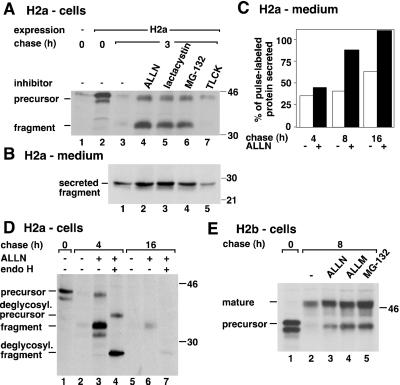
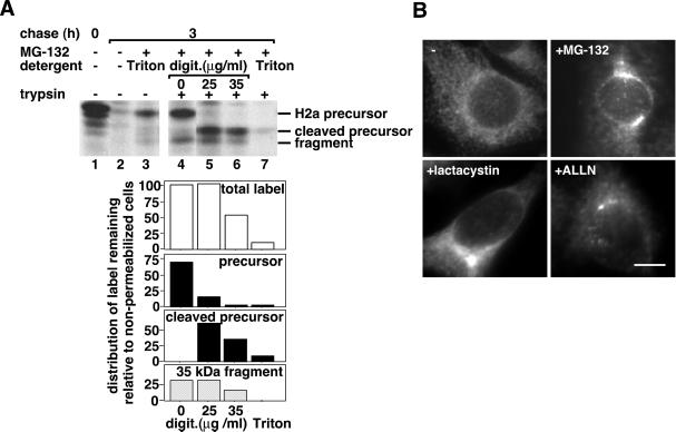
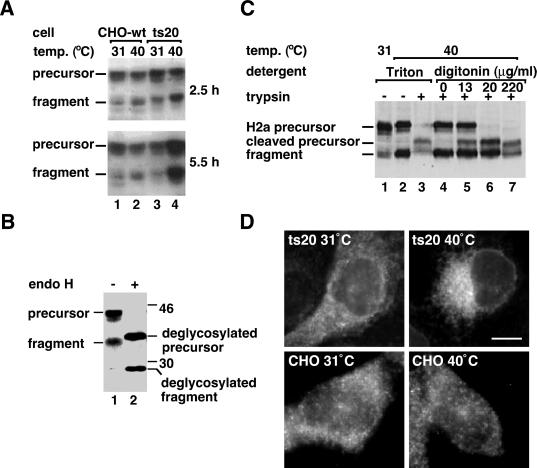
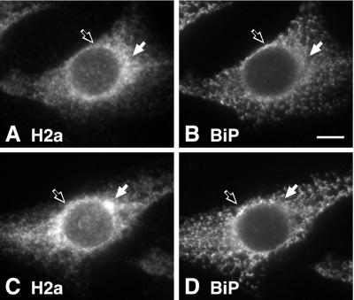
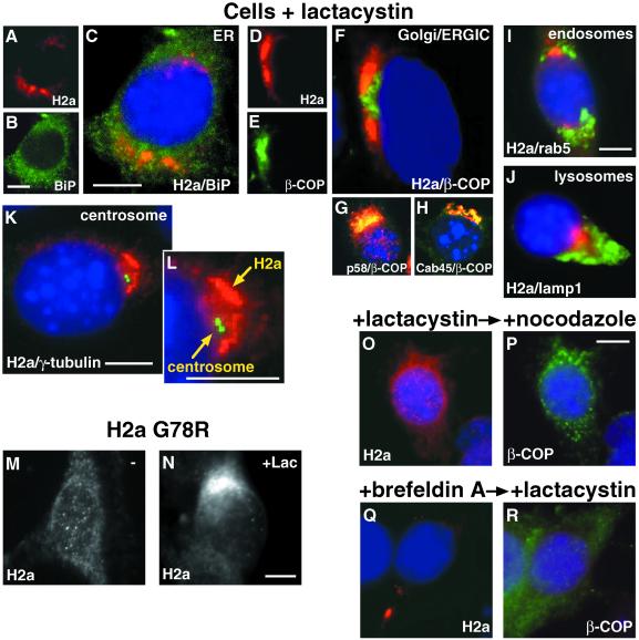
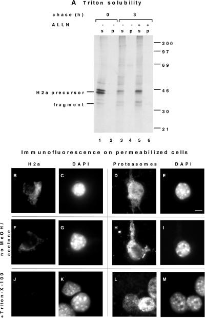
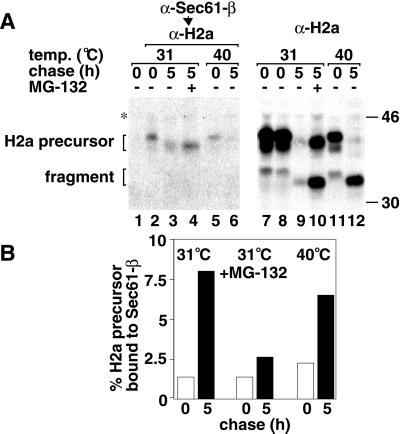
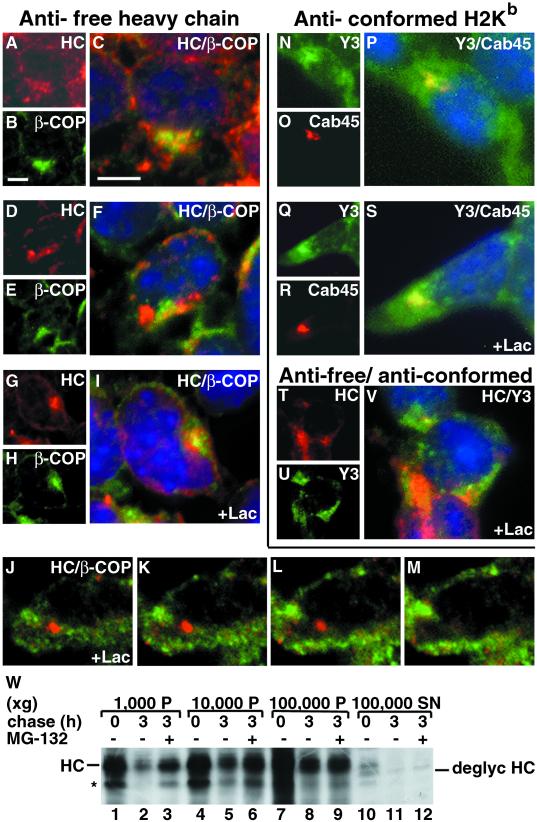
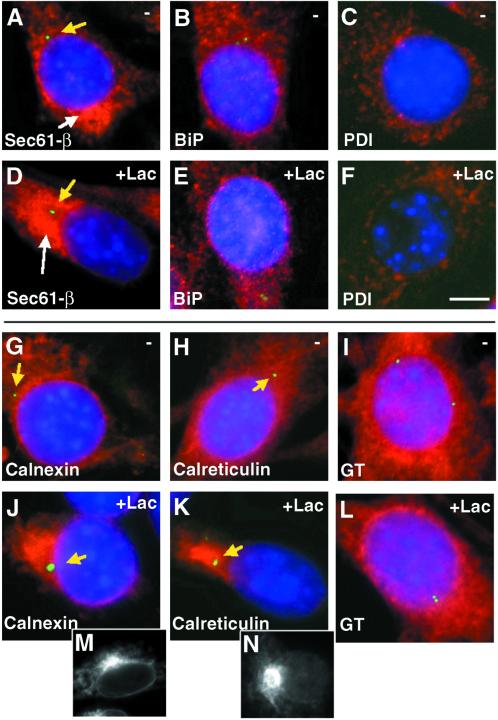
Degradation of proteins that, because of improper or suboptimal processing, are retained in the endoplasmic reticulum (ER) involves retrotranslocation to reach the cytosolic ubiquitin-proteasome machinery. We found that substrates of this pathway, the precursor of human asialoglycoprotein receptor H2a and free heavy chains of murine class I major histocompatibility complex (MHC), accumulate in a novel preGolgi compartment that is adjacent to but not overlapping with the centrosome, the Golgi complex, and the ER-to-Golgi intermediate compartment (ERGIC). On its way to degradation, H2a associated increasingly after synthesis with the ER translocon Sec61. Nevertheless, it remained in the secretory pathway upon proteasomal inhibition, suggesting that its retrotranslocation must be tightly coupled to the degradation process. In the presence of proteasomal inhibitors, the ER chaperones calreticulin and calnexin, but not BiP, PDI, or glycoprotein glucosyltransferase, concentrate in the subcellular region of the novel compartment. The "quality control" compartment is possibly a subcompartment of the ER. It depends on microtubules but is insensitive to brefeldin A. We discuss the possibility that it is also the site for concentration and retrotranslocation of proteins that, like the mutant cystic fibrosis transmembrane conductance regulator, are transported to the cytosol, where they form large aggregates, the "aggresomes."


Figures

Figure 1
Figure 1
Proteasomal inhibitors cause accumulation of ASGPR H2a membrane-bound precursor, its soluble 35-kDa fragment, and membrane-bound H2b inside the secretory pathway. (A) NIH 3T3 cells stably expressing H2a or untransfected (lane 1) were metabolically labeled with [35S] Cys for 20 min and chased with complete medium in the absence or in the presence of the indicated protease inhibitors. Incubations with the protease inhibitors were only during the chase period, with the following concentrations: 100 μM ALLN, ALLM, and TLCK, 25 μM lactacystin, and 10 μM MG-132. Cell lysates were immunoprecipitated, and immunoprecipitates were analyzed by SDS-PAGE followed by fluorography. On the left are indicated the positions for migration of H2a precursor and of its 35-kDa ectodomain fragment. On the right are molecular masses of protein standards in kilodaltons. (B) At the end of each chase period supernatants from cells in (A) were immunoprecipitated, and immunoprecipitates were treated with N-glycosidase F before SDS-PAGE and fluorography. The gel was exposed for 3 wk compared with 1 week in (A). (C) Phosphorimager quantitation of an experiment similar to that in (B). H2a ectodomain fragment secreted to the cell supernatants in the absence (white bars) or in the presence (black bars) of 100 μM ALLN were plotted as a percent of pulse-labeled H2a. (D) In a similar experiment to that in (A), the immunoprecipitates were treated in some cases with endo H before SDS-PAGE. (E) An experiment similar to that in (A) was done but with cells stably expressing H2b. On the left are indicated the positions for migration of H2b precursor and its mature Golgi-processed form.
Figure 2
Figure 2
H2a precursor and soluble ectodomain fragment accumulate in a membrane-enclosed compartment upon proteasomal inhibition. (A) After metabolic labeling similar to that in Figure 1 (A) and chase in the absence or presence of 10 μM MG-132, cells were incubated with or without trypsin in Triton X-100 or in low of H2aconcentrations of digitonin, as detailed in MATERIALS AND of H2a METHODS. Cleaved precursor of H2arefers to H2a precursor cleaved by trypsin. The lower panels are of H2aphosphorimager quantitations of this experiment, where the value of 100 represents the total amount of H2a after 3 h chase in the absence of digitonin (lane 4). (B) H2a immunofluorescence on fixed and permeabilized cells expressing H2a preincubated for 5 h with or without proteasome inhibitors at concentrations similar to those in Figure 1. Anti-H2 carboxyterminal were the primary antibodies and Cy3-conjugated goat antirabbit IgG the secondary. Bar = 10 μm.
Figure 3
Figure 3
Inhibition of ubiquitination causes H2a precursor and soluble ectodomain fragment to accumulate in a membrane-enclosed compartment. (A) A cell line carrying a thermosensitive mutant E1 (ts20 cells) and wild-type cells were both stably transfected with H2a cDNA. Representative clones of transfected cells were grown at 31°C and then incubated for the indicated times at 31°C (permissive temperature) or 40°C (restrictive), followed by lysis, SDS-PAGE, and immunoblotting with anti-H2 carboxyterminal antibodies. A secondary HRP-conjugated goat anti-rabbit IgG was used followed by ECL. (B) Ts20 cells expressing H2a were incubated for 8 h at the restrictive temperature, followed by lysis, treatment without (lane 1) or with endo H (lane 2), SDS-PAGE, and immunoblotting as in (A). (C) Ts20 cells expressing H2a were incubated for 5 h at the indicated temperatures. Before lysis cells were treated with Triton-X-100 or with low concentrations of digitonin in the presence or absence of trypsin as in Figure 2 A. SDS-PAGE and immunoblotting was as in (A). (D) H2a immunofluorescence as in Figure 2 B was visualized on ts20 cells expressing H2a incubated at 31°C or at 40°C for 5 h before fixation. Bar = 10 μm.
Figure 4
Figure 4
H2a is partially concentrated in a juxtanuclear region in some untreated cells. Cells expressing H2a were fixed and permeabilized, and double label immunofluorescence was performed with the use of anti-H2 carboxyterminal antibodies and Cy3-conjugated goat anti-rabbit IgG (A and C) and mouse anti-BiP antibodies with FITC-conjugated goat-anti-mouse IgG (B and D). A and B show a cell representative of the general population. C and D show a cell with an H2a immunofluorescence pattern representing ∼5% of the cells. The white arrows show regions with higher H2a concentration and not BiP and black outlined arrows show regions with higher BiP concentration and not H2a. Bar = 10 μm.
Figure 5
Figure 5
On proteasomal inhibition accumulated H2a is localized in a nocodazole-sensitive compartment, next to the centrosome, the Golgi and the ERGIC. Cells were incubated for 5 h with 25 μM lactacystin. Triple label immunofluorescence on fixed and permeabilized cells was done with secondary goat-antirabbit IgG conjugated to Cy3 with anti-H2a (panels A, C, D, F, I-O, Q), antip58 (ERGIC) (G) and anti-Cab45 (Golgi; Scherer et al., 1996) (H). FITC-conjugated goat-antimouse IgG was used with anti-BiP (B,C), anti-β-COP (E-H, P, R), antirab5 (I), and anti-γ-tubulin (K, L). FITC-conjugated goat-antirat IgG was used with antilamp1 (J). Nuclei were stained with DAPI. Overlap of Cy3 and FITC in panels G and H appears yellow. Immunofluorescence with anti-H2a antibodies was also done on fixed and permeabilized cells expressing the mutant H2a G78R (uncleavable; Yuk and Lodish, 1993; Neumann et al., 1996) (M) or on the same cells treated with lactacystin (Lac) (N). (O, P) Cells were treated for 3 h with 25 μM lactacystin with the addition of 20 μM nocodazole for an extra 2 h before fixation, permeabilization, and immunofluorescence (Q, R) Cells were treated for 2 h with 5 μg/ml brefeldin A, then with 25 μM lactacystin for an extra 3 h before fixation, permeabilization, and immunofluorescence. Bars = 10 μm.
Figure 6
Figure 6
H2a is not aggregated, and it is enclosed in juxtanuclear membranes, while proteasomes are on the cytosolic side. (A) Cells stably expressing H2a were metabolically labeled with [35S] Cys for 20 min and chased with complete medium for 3 h in the absence or in the presence of 100 μM ALLN. They were then lysed and immunoprecipitated as in Figure 1 (A), but immunoprecipitation was done also on material insoluble in Triton-containing buffer (“p”) and not only on the soluble (“s”) as described in MATERIALS AND METHODS. (B-E) Cells were treated for 5 h with 25 μM lactacystin. They were then permeabilized by breakage with a few strokes in a Dounce homogenizer, then adhered to coverslips with polylysine as described in MATERIALS AND METHODS. They were then fixed with methanol/acetone, and immunofluorescence was performed as in Figure 5, with the use of anti-H2a and DAPI (B, C) or antiproteasome antibody and DAPI (D, E). Cy3-conjugated secondary goat antirabbit IgG was used. Bar = 10 μm. (F-I) The same procedure was done as in (B-E), except that incubations with antibodies were done before fixation with methanol/acetone. (J-M) The same procedure was done as in (B-E), except that cells were treated with 0.2% Triton-X-100 before adhering to the coverslips.
Figure 7
Figure 7
H2a interacts with Sec61-β increasingly with chase time. (A) Ts20 cells expressing H2a were metabolically labeled, as in Figure 1 (A), at the indicated temperatures, in the absence or presence of 10 μM MG-132, solubilized with 1% digitonin in 20 mM HEPES, pH 7.6, and each sample was divided into 2 identical halves. These were immunoprecipitated with anti-Sec61-β antibodies (left panel) or with anti-H2a antibodies (right panel). Lane 1 is a control with preimmune antibodies instead of anti-Sec61-β. Immunoprecipitates in the right panel were run on SDS-PAGE, whereas those in the left panel were reimmunoprecipitated with anti-H2a after boiling in SDS containing buffer and addition of excess buffer A as described in Materials and Methods. The change in migration of H2a bands after chase is due to mannose trimming (Frenkel and Lederkremer, unpublished results). (B) Quotients of phosphorimager quantitation of H2a precursor bands in the left panel in (A) (Sec61-associated H2a) over those on the right panel (total H2a) were plotted for each treatment after pulse-labeling (white bars) or after 5 h chase (black bars).
Figure 8
Figure 8
Misfolded unassembled MHC class I heavy chains accumulate in a compartment, next to the Golgi and the ERGIC. (A) Triple label immunofluorescence on fixed and permeabilized A5O5 cells was done with secondary goat-antirabbit IgG conjugated to Cy3 with rabbit antimouse MHC class I free heavy chain antibodies (HC) (panels A, C, D, F, G, I-M, T, V) and anti-Cab45 (O, P, R, S). FITC-conjugated goat-antimouse IgG was used with anti-β-COP (B, C, E, F, H, I-M) and mouse antibodies against conformed assembled MHC class I H2Kb complexes (Y3) (N, P, Q, S, U, V). Nuclei were stained with DAPI in C, F, I, V. Cells in G-M and Q-V were incubated for 5 h with 25 μM lactacystin before fixation and immunofluorescence. Bars = 10 μm. (W) A5O5 cells were metabolically labeled with [35S] Met for 30 min and chased with complete medium in the absence or presence of 30 μM MG-132. Cells were then homogenized and fractionated, as detailed in MATERIALS AND METHODS. Lysates of each fraction pellet (P) or supernatant (SN) were immunoprecipitated with antifree heavy chain antibodies. Immunoprecipitates were analyzed by SDS-PAGE followed by fluorography. The bands corresponding to heavy chains (HC), deglycosylated heavy chains (deglyc HC) and a possible degradation product (*) are indicated.
Figure 9
Figure 9
Calnexin, calreticulin, and also partially Sec61-β accumulate next to the centrosome in the presence of lactacystin. Triple-label immunofluorescence (as in Figure 5) was performed on 3T3 cells expressing H2a preincubated without (A-C and G-I) or with 25 μM lactacystin (D-F and J-L), with the use of DAPI, FITC-conjugated goat antimouse IgG with anti-γ-tubulin, and Cy3-conjugated goat antirabbit IgG with anti-Sec61-β (A, D), anti-BiP (B, E), anti-PDI (C, F), anticalnexin (G, J), anticalreticulin (H, K), and anti-UDPGlc:glycoprotein glucosyltransferase (GT) (I, L). Bar = 10 μm. The yellow arrows point out the location of the centrosomes. Single label immunofluorescence is shown in the bottom panels for calnexin (M) and calreticulin (N) in cells incubated with lactacystin.

Similar articles

Cited by

References

    1. Amara JF, Lederkremer G, Lodish HF. Intracellular degradation of unassembled asialoglycoprotein receptor subunits: a pre-Golgi, nonlysosomal endoproteolytic cleavage. J Cell Biol. 1989;109:3315–3324. - PMC - PubMed
    1. Ayalon-Soffer M, Kamhi-Nesher S, Lederkremer GZ. Folding and self-assembly do not prevent ER retention and proteasomal degradation of asialoglycoprotein receptor H2a. FEBS Lett. 1999a;460:112–116. - PubMed
    1. Ayalon-Soffer M, Shenkman M, Lederkremer GZ. Differential role of mannose and glucose trimming in the ER degradation of asialoglycoprotein receptor subunits. J Cell Sci. 1999b;112:3309–3318. - PubMed
    1. Bebok Z, Mazzochi C, King SA, Hong JS, Sorscher EJ. The mechanism underlying cystic fibrosis transmembrane conductance regulator transport from the endoplasmic reticulum to the proteasome includes Sec61β and a cytosolic, deglycosylated intermediary. J Biol Chem. 1998;273:29873–29878. - PubMed
    1. Biederer T, Volkwein C, Sommer T. Role of Cue1p in ubiquitination and degradation at the ER surface. Science. 1997;278:1806–1809. - PubMed

Publication types

MeSH terms